Call us
301-363-4651 (Available 9 a.m. to 5 p.m. CST from Monday to Friday)
| Code | CSB-RA188887A0HU |
| Size | US$210 |
| Order now | |
| Image |
|
| Have Questions? | Leave a Message or Start an on-line Chat |
| Application | Recommended Dilution |
|---|---|
| WB | 1:500-1:2000 |
| IF | 1:50-1:200 |
| FC | 1:50-1:200 |
Glutathione synthetase catalyzes the final step in glutathione biosynthesis, converting gamma-glutamylcysteine and glycine into the essential antioxidant glutathione. This enzyme plays a central role in cellular defense against oxidative stress, xenobiotic detoxification, and maintenance of redox homeostasis. Researchers investigating metabolic disorders, oxidative damage pathways, and cancer metabolism frequently require reliable tools to monitor GSS expression across experimental models.
This recombinant monoclonal antibody, generated from clone 2H9 against a synthetic peptide derived from human GSS, offers the consistency and reproducibility that demanding research protocols require. Because recombinant antibodies are produced from defined sequences rather than traditional hybridoma methods, you can expect lot-to-lot uniformity that supports longitudinal studies and reproducible data across your research program.
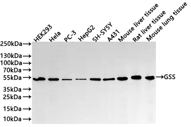
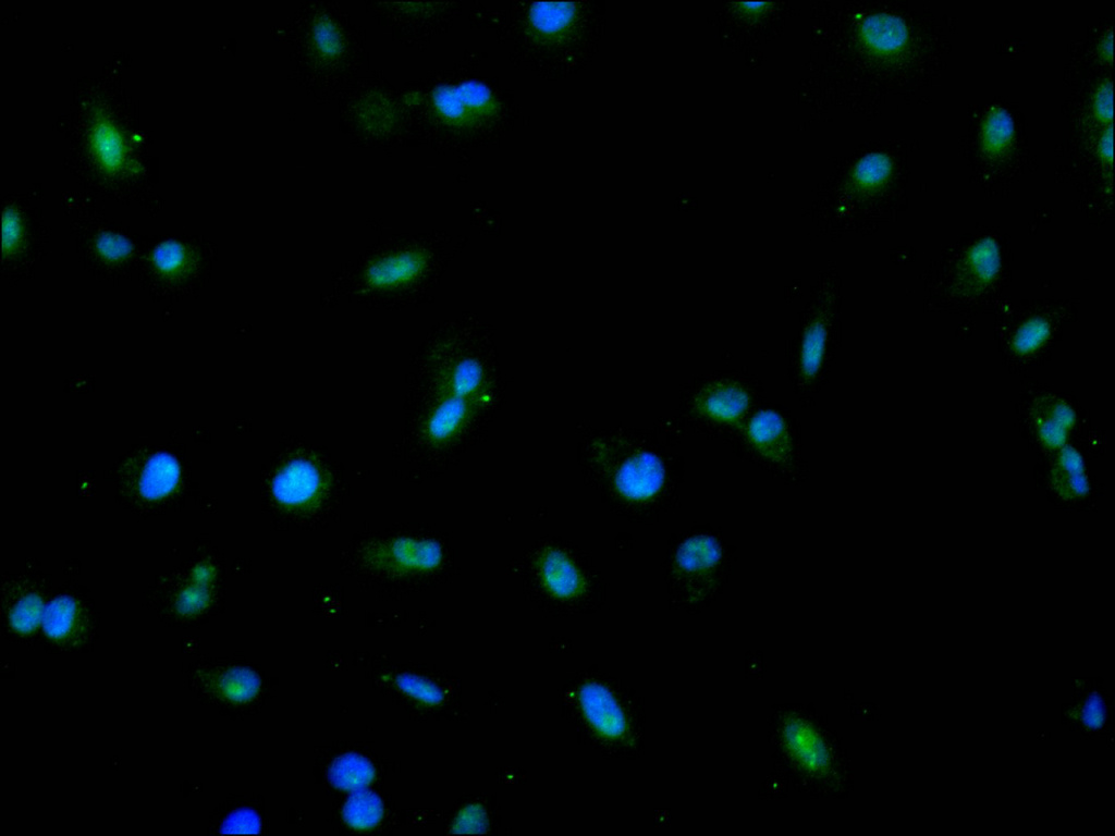
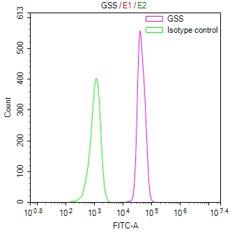
Validation testing demonstrates robust performance across multiple experimental platforms. Western blot analysis detects a clean band at the predicted 52 kDa molecular weight across diverse human cell lines including HEK293, HeLa, PC-3, HepG2, SH-SY5Y, and A431 cells at dilutions ranging from 1:500 to 1:2000. Cross-species reactivity extends to mouse and rat samples, with confirmed detection in mouse liver and lung tissue as well as rat liver tissue, providing flexibility for translational studies using rodent models. Immunofluorescence staining in A549 cells reveals cytoplasmic localization patterns consistent with the enzyme's known subcellular distribution, while flow cytometry analysis in A431 cells demonstrates clear separation from isotype controls.
Whether you are characterizing glutathione metabolism in cancer cell lines, investigating oxidative stress responses in tissue samples, or examining GSS expression across species, this antibody provides the validated performance and application versatility to support your experimental objectives.
There are currently no reviews for this product.

