Call us
301-363-4651 (Available 9 a.m. to 5 p.m. CST from Monday to Friday)
| Code | CSB-RA072684A0HU |
| Size | US$210 |
| Order now | |
| Image |
|
| Have Questions? | Leave a Message or Start an on-line Chat |
| Application | Recommended Dilution |
|---|---|
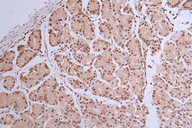
| IHC | 1:50-1:200 |
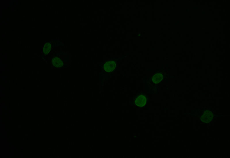
| IF | 1:50-1:200 |
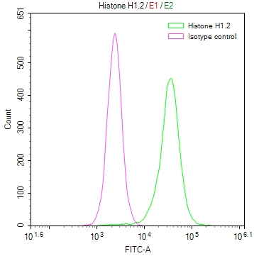
| FC | 1:50-1:200 |
The HIST1H1C recombinant monoclonal antibody is a product of a carefully planned production process. It commences with in vitro cloning, where the HIST1H1C antibody genes are seamlessly integrated into expression vectors. Subsequently, these vectors are introduced into host cells, paving the way for the recombinant antibody's expression within a cell culture setting. Post-expression, the HIST1H1C recombinant monoclonal antibody undergoes purification from the supernatant of transfected host cell lines through affinity chromatography. This antibody shows a specific binding affinity for the human HIST1H1C protein in ELISA, IHC, IF, and FC applications.
HIST1H1C, also called Histone H1.2, like other histone proteins, plays a significant role in the packaging and organization of DNA within the cell nucleus. Specifically, its main function is related to chromatin compaction and gene regulation.
There are currently no reviews for this product.

