| Image |
-
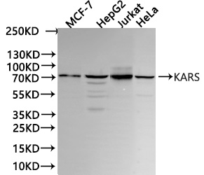
Western Blot
Positive WB detected in: MCF-7 whole cell lysate(30µg), HepG2 whole cell lysate(30µg), Jurkat whole cell lysate(30µg), HeLa whole cell lysate(30µg)
All lanes: KARS antibody at 1:1000
Secondary
Goat polyclonal to Human IgG at 1/40000 dilution
Predicted band size: 68 kDa
Observed band size:72 kDa
Exposure time: 15S
-
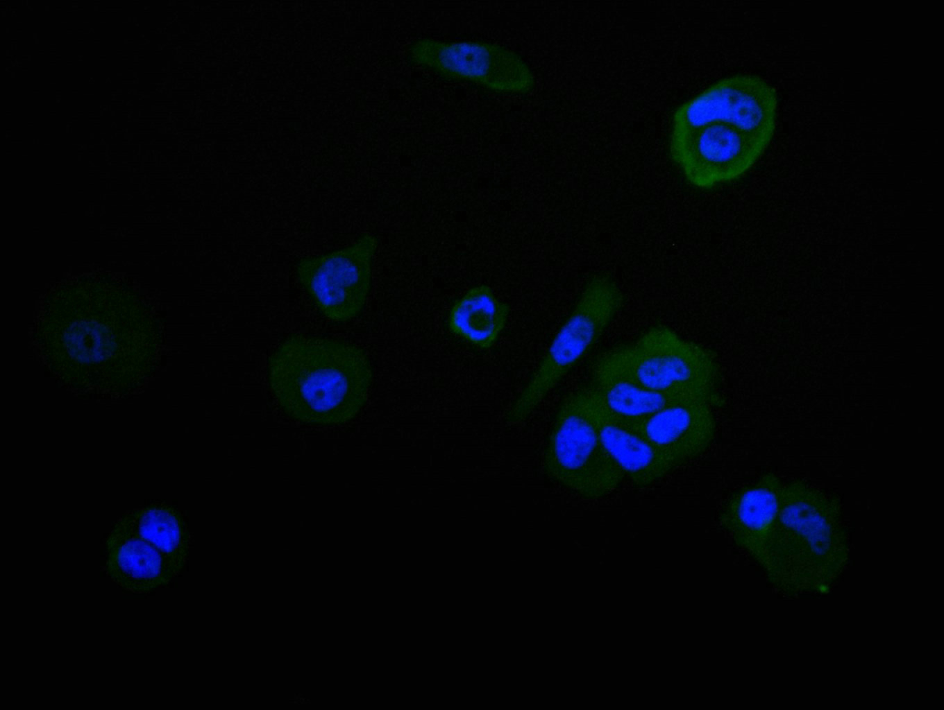
Immunofluorescence staining of MCF-7 cell with CSB-RA618973MA1HU at 1:30, counter-stained with DAPI. The cells were fixed in 4% formaldehyde, permeabilized using 0.2% Triton X-100 and blocked in 10% normal Goat Serum. The cells were then incubated with the antibody overnight at 4°C. The secondary antibody was Fluorescein (FITC) AffiniPure Goat Anti-Human IgG, Fcγ fragment specific.
-
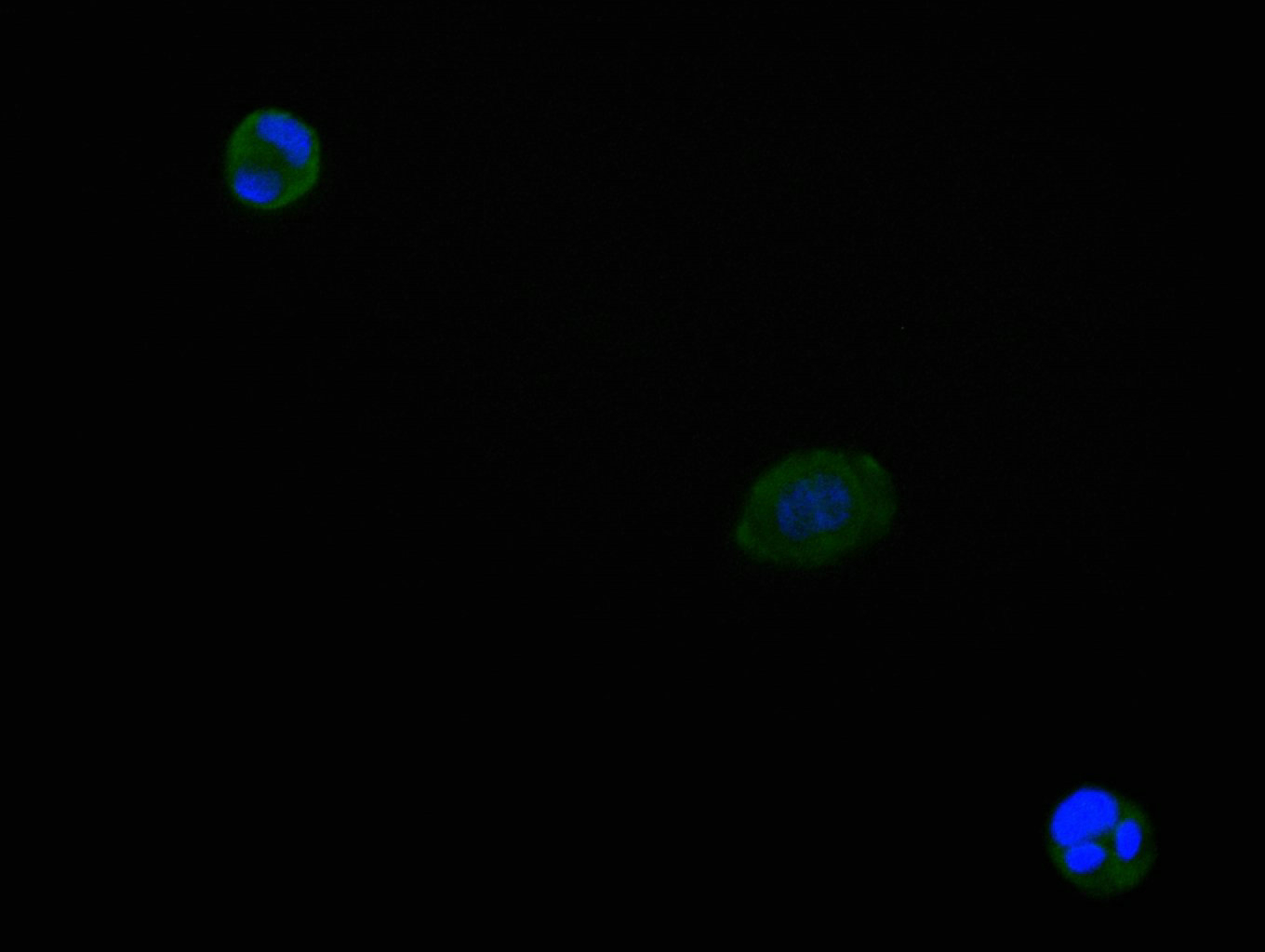
Immunofluorescence staining of CACO-2 cell with CSB-RA618973MA1HU at 1:30, counter-stained with DAPI. The cells were fixed in 4% formaldehyde, permeabilized using 0.2% Triton X-100 and blocked in 10% normal Goat Serum. The cells were then incubated with the antibody overnight at 4°C. The secondary antibody was Fluorescein (FITC) AffiniPure Goat Anti-Human IgG, Fcγ fragment specific.
-
Overlay Peak curve showing Raji cells stained with CSB-RA618973MA1HU (red line) at 1:100. The cells were fixed in 4% formaldehyde and permeated by 0.2% TritonX-100 for 10min. Then 10% normal goat serum to block non-specific protein-protein interactions followed by the antibody (1ug/1*106cells) for 45min at 4℃. The secondary antibody used was Fluorescein (FITC) AffiniPure Goat Anti-Human IgG, Fcγ fragment specific at 1:200 dilution for 35 min at 4℃.Control antibody (green line) was human IgG1 (1ug/1*106cells) used under the same conditions. Acquisition of >10, 000 events was performed.
|