MAPRE3 Recombinant Monoclonal Antibody

Code CSB-RA073454A0HU
Size US$210
Order now
Image
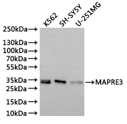
  • Western Blot
    Positive WB detected in: K562 whole cell lysate(30µg), SH-SY5Y whole cell lysate(30µg), U-251MG whole cell lysate(30µg)
    All lanes: MAPRE3 antibody at 1:1000
    Secondary
    Goat polyclonal to rabbit IgG at 1/40000 dilution
    Predicted band size: 32 kDa
    Observed band size: 32 kDa
    Exposure time:2min
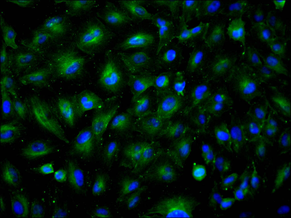
  • Immunofluorescence staining of U-251MG cell with CSB-RA073454A0HU at 1:50, counter-stained with DAPI. The cells were fixed in 4% formaldehyde, permeabilized using 0.2% Triton X-100 and blocked in 10% normal Goat Serum. The cells were then incubated with the antibody overnight at 4°C. The secondary antibody was Alexa Fluor 488-congugated AffiniPure Goat Anti-Rabbit IgG(H+L).
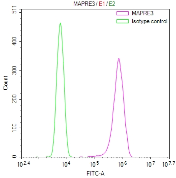
  • Overlay Peak curve showing SH-SY5Y cells stained with CSB-RA073454A0HU (red line) at 1:100. The cells were fixed in 4% formaldehyde and permeated by 0.2% TritonX-100 for 10min. Then 10% normal goat serum to block non-specific protein-protein interactions followed by the antibody (1ug/1*106cells) for 45min at 4℃. The secondary antibody used was FITC-conjugated goat anti-rabbit IgG (H+L) at 1/200 dilution for 35min at 4℃.Control antibody (green line) was Rabbit IgG (1ug/1*106cells) used under the same conditions. Acquisition of >10,000 events was performed.
Have Questions? Leave a Message or Start an on-line Chat

Product Details

Uniprot No.
Target Names
MAPRE3
Alternative Names
Microtubule-associated protein RP/EB family member 3 (EB1 protein family member 3) (EBF3) (End-binding protein 3) (EB3) (RP3), MAPRE3
Species Reactivity
Human
Immunogen
Recombinant Antibody
Immunogen Species
Homo sapiens (Human)
Conjugate
Non-conjugated
Clonality
Monoclonal
Isotype
Rabbit IgG
Clone No.
8H1
Purification Method
Affinity-chromatography
Concentration
It differs from different batches. Please contact us to confirm it.
Buffer
Rabbit IgG in 10mM phosphate buffered saline , pH 7.4, 150mM sodium chloride, 0.05% BSA, 0.02% sodium azide and 50% glycerol.
Form
Liquid
Tested Applications
ELISA, WB, IF, FC
Recommended Dilution
Application Recommended Dilution
WB 1:500-1:5000
IF 1:50-1:200
FC 1:50-1:200
Troubleshooting and FAQs
Storage
Upon receipt, store at -20°C or -80°C. Avoid repeated freeze.
Lead Time
Basically, we can dispatch the products out in 1-3 working days after receiving your orders. Delivery time maybe differs from different purchasing way or location, please kindly consult your local distributors for specific delivery time.
Description

MAPRE3, also known as End-binding protein 3 (EB3), plays a critical role in microtubule dynamics by tracking the plus ends of growing microtubules. As a member of the RP/EB protein family, MAPRE3 regulates microtubule stability and is involved in essential cellular processes including mitosis, cell migration, and neuronal development. Its dynamic localization patterns make it a valuable marker for studying cytoskeletal organization and intracellular transport mechanisms.

This recombinant monoclonal antibody, generated against a synthetic peptide derived from human MAPRE3, offers the consistency and reliability that demanding experimental workflows require. As a sequence-defined reagent produced through recombinant technology, it eliminates the lot-to-lot variability inherent in traditional hybridoma-derived antibodies, ensuring reproducible results across long-term studies and multi-site collaborations.

Validation studies demonstrate robust performance across multiple detection platforms. Western blot analysis confirms specific detection of MAPRE3 at the expected 32 kDa molecular weight in K562, SH-SY5Y neuroblastoma, and U-251MG glioblastoma cell lysates, providing confidence when working with diverse human cell models. Immunofluorescence staining in U-251MG cells reveals clear cytoplasmic localization patterns consistent with MAPRE3's known association with microtubule structures. Flow cytometry validation in SH-SY5Y cells further demonstrates the antibody's utility for quantitative single-cell analysis of intracellular targets.

This versatility across immunofluorescence and flow cytometry applications at working dilutions of 1:50-1:200 makes this antibody particularly well-suited for researchers investigating microtubule dynamics, cytoskeletal regulation, and neuronal cell biology. Its validated performance in neuroblastoma and glioblastoma cell lines positions it as a valuable tool for cancer research focused on cytoskeletal alterations.

Usage
For Research Use Only. Not for use in diagnostic or therapeutic procedures.

Customer Reviews and Q&A

 Customer Reviews

There are currently no reviews for this product.

Submit a Review here

Target Background

Function
Plus-end tracking protein (+TIP) that binds to the plus-end of microtubules and regulates the dynamics of the microtubule cytoskeleton. Promotes microtubule growth. May be involved in spindle function by stabilizing microtubules and anchoring them at centrosomes. Also acts as a regulator of minus-end microtubule organization: interacts with the complex formed by AKAP9 and PDE4DIP, leading to recruit CAMSAP2 to the Golgi apparatus, thereby tethering non-centrosomal minus-end microtubules to the Golgi, an important step for polarized cell movement. Promotes elongation of CAMSAP2-decorated microtubule stretches on the minus-end of microtubules. May play a role in cell migration.
Gene References into Functions
  1. One candidate pathway coupling actin filaments to microtubules consists of the actin filament-binding protein drebrin and the microtubule-binding +TIP protein EB3. This pathway is regulated proximally by cyclin-dependent kinase 5 phosphorylation of drebrin but the upstream elements in the pathway have yet to be identified. PMID: 28865014
  2. Findings show that co-ordination of dynamic microtubules and actin filaments by the drebrin/EB3 pathway drives prostate cancer cell invasion and is therefore implicated in disease progression. PMID: 28319065
  3. Findings suggest that methylation-associated down-regulation of EBF3 and IRX1 genes may play an important role in a pathogenic effect of TGF-beta on RASFs. PMID: 23456299
  4. Daughter cell adhesion and cytokinesis completion are spatially regulated by distinct states of EB3 phosphorylation on serine 176 by Aurora B. PMID: 23712260
  5. decreasing the drebrin E levels disrupted the normal subapical F-actin-myosin-IIB-betaII-spectrin network and the apical accumulation of EB3, a microtubule-plus-end-binding protein PMID: 22275434
  6. VE-cadherin outside-in signaling regulates cytosolic calcium homeostasis and EB3 phosphorylation. PMID: 23159740
  7. study found a polycytosine repeat (C8) in exon 5 of MAPRE3 that could be a potential mutation target in cancers with microsatellite instability (MSI); found that the C8 is frequently mutated in gastric and colorectal carcinomas with MSI PMID: 20632835
  8. The RP3 protein is involved in SATB1-mediated gene repression of Bcl2 and dynein subunit rp3 has functions independent of the dynein motor. PMID: 16079286
  9. Data show that two mitotic kinases, Aurora-A and Aurora-B, phosphorylate endogenous EB3 at Ser-176, and the phosphorylation triggers disruption of the EB3-SIAH-1 complex, resulting in EB3 stabilization during mitosis. PMID: 19696028

Show More

Hide All

Subcellular Location
Cytoplasm, cytoskeleton.
Protein Families
MAPRE family
Tissue Specificity
Predominantly expressed in brain and muscle.
Database Links

HGNC: 6892

OMIM: 605788

KEGG: hsa:22924

STRING: 9606.ENSP00000233121

UniGene: Hs.515860

icon of phone
Call us
301-363-4651 (Available 9 a.m. to 5 p.m. CST from Monday to Friday)

icon of address
Address
No. 269, Shendun 5th Road, Donghu Hi-Tech Development Area, Hubei Province, 430206, P.R.China
icon of social media
Join us with

Subscribe newsletter

Leave a message

* To protect against spam, please pass the CAPTCHA test below.
CAPTCHA verification
© 2007-2026 CUSABIO TECHNOLOGY LLC All rights reserved. 鄂ICP备15011166号-1
Place an order now

I. Product details

*
*
*
*

II. Contact details

*
*

III. Ship To

*
*
*
*
*
*
*

IV. Bill To

*
*
*
*
*
*
*
*