| Image |
-
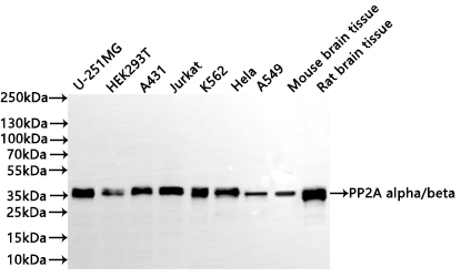
Western Blot
Positive WB detected in: U-251MG whole cell lysate(30µg), HEK293T whole cell lysate(30µg), A431 whole cell lysate(30µg), Jurkat whole cell lysate(30µg), K562 whole cell lysate(30µg), Hela whole cell lysate(30µg), A549 whole cell lysate(30µg), Mouse brain tissue lysate(30µg), Rat brain tissue lysate(30µg)
All lanes: PP2A alpha/beta antibody at 1:1000
Secondary
Goat polyclonal to rabbit IgG at 1/40000 dilution
Predicted band size: 36 kDa
Observed band size: 36 kDa
Exposure time:5s
-
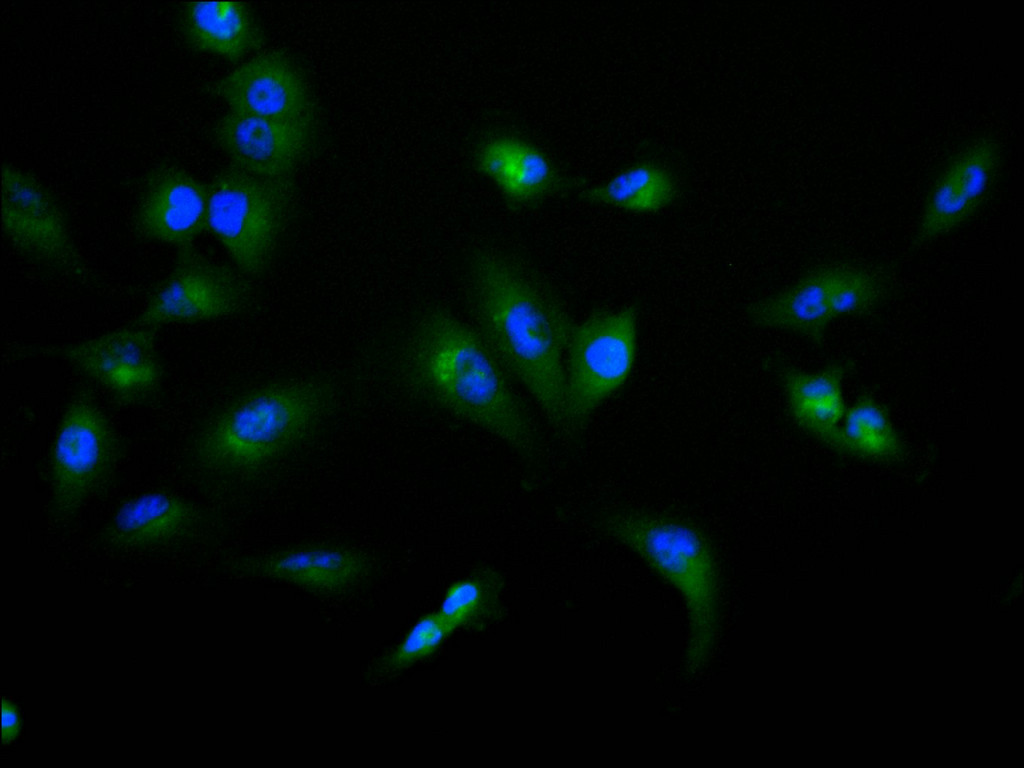
Immunofluorescence staining of U-251MG cell with CSB-RA616740A0HU at 1:50, counter-stained with DAPI. The cells were fixed in 4% formaldehyde, permeabilized using 0.2% Triton X-100 and blocked in 10% normal Goat Serum. The cells were then incubated with the antibody overnight at 4°C. The secondary antibody was Alexa Fluor 488-congugated AffiniPure Goat Anti-Rabbit IgG(H+L).
-
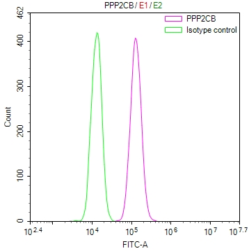
Overlay Peak curve showing 786-O cells stained with CSB-RA616740A0HU (red line) at 1:100. The cells were fixed in 4% formaldehyde and permeated by 0.2% TritonX-100 for 10min. Then 10% normal goat serum to block non-specific protein-protein interactions followed by the antibody (1ug/1*106cells) for 45min at 4℃. The secondary antibody used was FITC-conjugated goat anti-rabbit IgG (H+L) at 1/200 dilution for 35min at 4℃.Control antibody (green line) was Rabbit IgG (1ug/1*106cells) used under the same conditions. Acquisition of >10,000 events was performed.
|