SEPTIN8 Recombinant Monoclonal Antibody

Code CSB-RA831681A0HU
Size US$210
Order now
Image
  • Western Blot
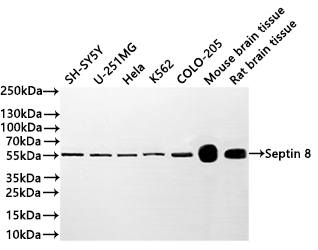
    Positive WB detected in: SH-SY5Y whole cell lysate(20µg),U-251MG whole cell lysate(20µg), Hela whole cell lysate(20µg), K562 whole cell lysate(20µg),COLO-205 whole cell lysate(20µg), Mouse brain tissue lysate(20µg), Rat brain tissue lysate(20µg)
    All lanes: Septin 8 antibody at 1:1000
    Secondary
    Goat polyclonal to rabbit IgG at 1/40000 dilution
    Predicted band size: 56 kDa
    Observed band size: 56 kDa
    Exposure time:2min
  • IHC image of CSB-RA831681A0HU diluted at 1:100 and staining in paraffin-embedded human stomach tissue performed on a Leica BondTM system. After dewaxing and hydration, antigen retrieval was mediated by high pressure in a citrate buffer (pH 6.0). Section was blocked with 10% normal goat serum 30min at RT. Then primary antibody (1% BSA) was incubated at 4°C overnight. The primary is detected by a Goat anti-rabbit polymer IgG labeled by HRP and visualized using 0.05% DAB.
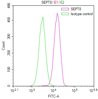
  • Overlay Peak curve showing 786-O cells stained with CSB-RA831681A0HU (red line) at 1:100. The cells were fixed in 4% formaldehyde and permeated by 0.2% TritonX-100 for 10min. Then 10% normal goat serum to block non-specific protein-protein interactions followed by the antibody (1ug/1*106cells) for 45min at 4℃. The secondary antibody used was FITC-conjugated goat anti-rabbit IgG (H+L) at 1/200 dilution for 35min at 4℃.Control antibody (green line) was Rabbit IgG (1ug/1*106cells) used under the same conditions. Acquisition of >10,000 events was performed.
Have Questions? Leave a Message or Start an on-line Chat

Product Details

Uniprot No.
Target Names
SEPTIN8
Alternative Names
SEPTIN8 antibody; KIAA0202 antibody; SEPT8 antibody; Septin-8 antibody
Species Reactivity
Human, Mouse, Rat
Immunogen
A synthesized peptide derived from human SEPTIN8
Immunogen Species
Homo sapiens (Human)
Conjugate
Non-conjugated
Clonality
Monoclonal
Isotype
Rabbit IgG
Clone No.
17B12
Purification Method
Affinity-chromatography
Concentration
It differs from different batches. Please contact us to confirm it.
Buffer
Rabbit IgG in 10mM phosphate buffered saline , pH 7.4, 150mM sodium chloride, 0.05% BSA, 0.02% sodium azide and 50% glycerol.
Form
Liquid
Tested Applications
ELISA, WB, IHC, FC
Recommended Dilution
Application Recommended Dilution
WB 1:500-1:2000
IHC 1:50-1:200
FC 1:50-1:200
Troubleshooting and FAQs
Storage
Upon receipt, store at -20°C or -80°C. Avoid repeated freeze.
Lead Time
Basically, we can dispatch the products out in 1-3 working days after receiving your orders. Delivery time maybe differs from different purchasing way or location, please kindly consult your local distributors for specific delivery time.
Description

Septin-8 belongs to the septin family of GTP-binding proteins that play essential roles in cytokinesis, membrane dynamics, and cytoskeletal organization. As a component of septin filament complexes, SEPTIN8 contributes to cellular compartmentalization and has been implicated in neurological function, making it a valuable target for researchers investigating cytoskeletal biology, cell division mechanisms, and neurodegenerative disease pathways.

This recombinant monoclonal antibody offers the reproducibility and consistency that demanding experimental workflows require. Generated from a defined clone (17B12) and produced using recombinant technology, it eliminates the lot-to-lot variability associated with traditional hybridoma-derived antibodies, ensuring your results remain comparable across extended studies and collaborative projects.

Validation data demonstrates robust performance across multiple applications and sample types. Western blot analysis confirms detection of the expected 56 kDa band in diverse human cell lines including SH-SY5Y neuroblastoma, U-251MG glioblastoma, HeLa, K562, and COLO-205 cells, providing flexibility for various experimental models. Cross-species reactivity extends to mouse and rat brain tissue lysates, supporting translational research using rodent models. Immunohistochemistry validation in paraffin-embedded human stomach tissue confirms utility for tissue-based studies, while flow cytometry analysis in 786-O cells demonstrates clear positive signal separation, enabling quantitative single-cell investigations.

The antibody's cross-species reactivity with human, mouse, and rat samples makes it particularly well-suited for comparative studies and preclinical research. Whether investigating septin dynamics in cancer cell biology, exploring neuronal cytoskeletal architecture, or examining cell division processes, this antibody provides a reliable tool for advancing your understanding of septin-mediated cellular functions.

Usage
For Research Use Only. Not for use in diagnostic or therapeutic procedures.

Customer Reviews and Q&A

 Customer Reviews

There are currently no reviews for this product.

Submit a Review here

Target Background

Function
Filament-forming cytoskeletal GTPase. May play a role in platelet secretion. Seems to participate in the process of SNARE complex formation in synaptic vesicles.; Stabilizes BACE1 protein levels and promotes the sorting and accumulation of BACE1 to the recycling or endosomal compartments, modulating the beta-amyloidogenic processing of APP.
Gene References into Functions
  1. SEPT8 modulates beta-amyloidogenic processing of APP through a mechanism affecting the intracellular sorting and accumulation of BACE1. PMID: 27084579
  2. Cloning, overexpression, purification and preliminary characterization of human septin 8. Fluorescence emission spectra, analytic gel filtration and DLS reflect the sample oligomeric heterogeneity with the predominance of dimers in solution. PMID: 20544379
  3. SEPT8 and SEPT4 are localized surrounding alpha-granules. Activation of platelets by agonists resulted in the translocation of SEPT4 and SEPT8 to the platelet surface indicating a possible functional role of these proteins in platelet granular secretion PMID: 15116257
  4. We studied the assembly of three human septins, SEPT4, SEPT5 and SEPT8, with each other (heterotypic) and with themselves (homotypic) using a yeast two-hybrid system. PMID: 15214843
  5. Our finding suggests a role for members of the septin family in the development of proliferative retinal membranes. PMID: 17625225

Show More

Hide All

Subcellular Location
Cytoplasm. Cytoplasm, cytoskeleton. Cell junction, synapse. Cell projection, axon. Cytoplasmic vesicle, secretory vesicle, synaptic vesicle membrane. Cell junction, synapse, presynapse.
Protein Families
TRAFAC class TrmE-Era-EngA-EngB-Septin-like GTPase superfamily, Septin GTPase family
Tissue Specificity
Widely expressed, including in brain, heart and platelets; most abundant in aorta. Isoform 2 is expressed at low levels in specific brain areas, such as occipital pole, frontal lobe, temporal lobe and putamen. Isoform 1 and 3 are highly expressed in speci
Database Links

HGNC: 16511

OMIM: 608418

KEGG: hsa:23176

STRING: 9606.ENSP00000367991

UniGene: Hs.522057

icon of phone
Call us
301-363-4651 (Available 9 a.m. to 5 p.m. CST from Monday to Friday)

icon of address
Address
No. 269, Shendun 5th Road, Donghu Hi-Tech Development Area, Hubei Province, 430206, P.R.China
icon of social media
Join us with

Subscribe newsletter

Leave a message

* To protect against spam, please pass the CAPTCHA test below.
CAPTCHA verification
© 2007-2026 CUSABIO TECHNOLOGY LLC All rights reserved. 鄂ICP备15011166号-1
Place an order now

I. Product details

*
*
*
*

II. Contact details

*
*

III. Ship To

*
*
*
*
*
*
*

IV. Bill To

*
*
*
*
*
*
*
*