UQCRFS1 Recombinant Monoclonal Antibody

Code CSB-RA928009A0HU
Size US$210
Order now
Image
  • Western Blot
    Positive WB detected in: MCF-7 whole cell lysate(20µg), U937 whole cell lysate(20µg),Hela whole cell lysate(20µg),HEK293T whole cell lysate(20µg),HL-60 whole cell lysate(20µg),HepG2 whole cell lysate(20µg), Mouse heart tissue lysate(20µg), Mouse liver tissue lysate(20µg)
    All lanes: UQCRFS1 antibody at 1:1000
    Secondary
    Goat polyclonal to rabbit IgG at 1/40000 dilution
    Predicted band size: 23/30 kDa
    Observed band size: 23 kDa
    Exposure time:2min
  • IHC image of CSB-RA928009A0HU diluted at 1:100 and staining in paraffin-embedded human breast cancer performed on a Leica BondTM system. After dewaxing and hydration, antigen retrieval was mediated by high pressure in a citrate buffer (pH 6.0). Section was blocked with 10% normal goat serum 30min at RT. Then primary antibody (1% BSA) was incubated at 4°C overnight. The primary is detected by a Goat anti-rabbit polymer IgG labeled by HRP and visualized using 0.05% DAB.
  • IHC image of CSB-RA928009A0HU diluted at 1:100 and staining in paraffin-embedded human stomach tissue performed on a Leica BondTM system. After dewaxing and hydration, antigen retrieval was mediated by high pressure in a citrate buffer (pH 6.0). Section was blocked with 10% normal goat serum 30min at RT. Then primary antibody (1% BSA) was incubated at 4°C overnight. The primary is detected by a Goat anti-rabbit polymer IgG labeled by HRP and visualized using 0.05% DAB.
  • Overlay Peak curve showing MCF-7 cells stained with CSB-RA928009A0HU (red line) at 1:100. The cells were fixed in 4% formaldehyde and permeated by 0.2% TritonX-100 for 10min. Then 10% normal goat serum to block non-specific protein-protein interactions followed by the antibody (1ug/1*106cells) for 45min at 4℃. The secondary antibody used was FITC-conjugated goat anti-rabbit IgG (H+L) at 1/200 dilution for 35min at 4℃.Control antibody (green line) was Rabbit IgG (1ug/1*106cells) used under the same conditions. Acquisition of >10,000 events was performed.
Have Questions? Leave a Message or Start an on-line Chat

Product Details

Uniprot No.
Target Names
Alternative Names
Complex III subunit 5 antibody; Complex III subunit IX antibody; Cytochrome b c1 complex subunit Rieske mitochondrial antibody; Cytochrome b-c1 complex subunit 11 antibody; Cytochrome b-c1 complex subunit 5 antibody; Cytochrome b6f complex iron sulfur subunit, chloroplastic antibody; petC antibody; PGR1 antibody; Plastohydroquinone:plastocyanin oxidoreductase iron sulfur protein antibody; Proton gradient regulation protein 1 antibody; Rieske iron sulfur protein antibody; Rieske iron-sulfur protein antibody; RIP1 antibody; RIS1 antibody; RISP antibody; Ubiquinol cytochrome C reductase rieske iron sulphur antibody; Ubiquinol cytochrome c reductase Rieske iron-sulfur polypeptide 1 antibody; Ubiquinol-cytochrome c reductase 8 kDa protein antibody; Ubiquinol-cytochrome c reductase iron-sulfur subunit antibody; UCRI_HUMAN antibody; UQCR5 antibody; UQCRFS1 antibody
Species Reactivity
Human, Mouse
Immunogen
A synthesized peptide derived from human UQCRFS1
Immunogen Species
Homo sapiens (Human)
Conjugate
Non-conjugated
Clonality
Monoclonal
Isotype
Rabbit IgG
Clone No.
4F10
Purification Method
Affinity-chromatography
Concentration
It differs from different batches. Please contact us to confirm it.
Buffer
Rabbit IgG in 10mM phosphate buffered saline , pH 7.4, 150mM sodium chloride, 0.05% BSA, 0.02% sodium azide and 50% glycerol.
Form
Liquid
Tested Applications
ELISA, WB, IHC, FC
Recommended Dilution
Application Recommended Dilution
WB 1:500-1:2000
IHC 1:50-1:200
FC 1:50-1:200
Troubleshooting and FAQs
Storage
Upon receipt, store at -20°C or -80°C. Avoid repeated freeze.
Lead Time
Basically, we can dispatch the products out in 1-3 working days after receiving your orders. Delivery time maybe differs from different purchasing way or location, please kindly consult your local distributors for specific delivery time.
Usage
For Research Use Only. Not for use in diagnostic or therapeutic procedures.

Customer Reviews and Q&A

 Customer Reviews

There are currently no reviews for this product.

Submit a Review here

Target Background

Function
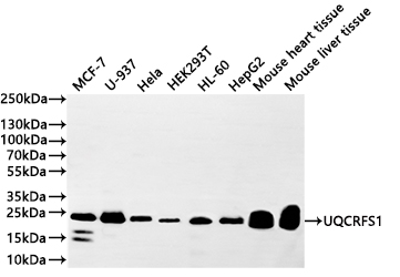
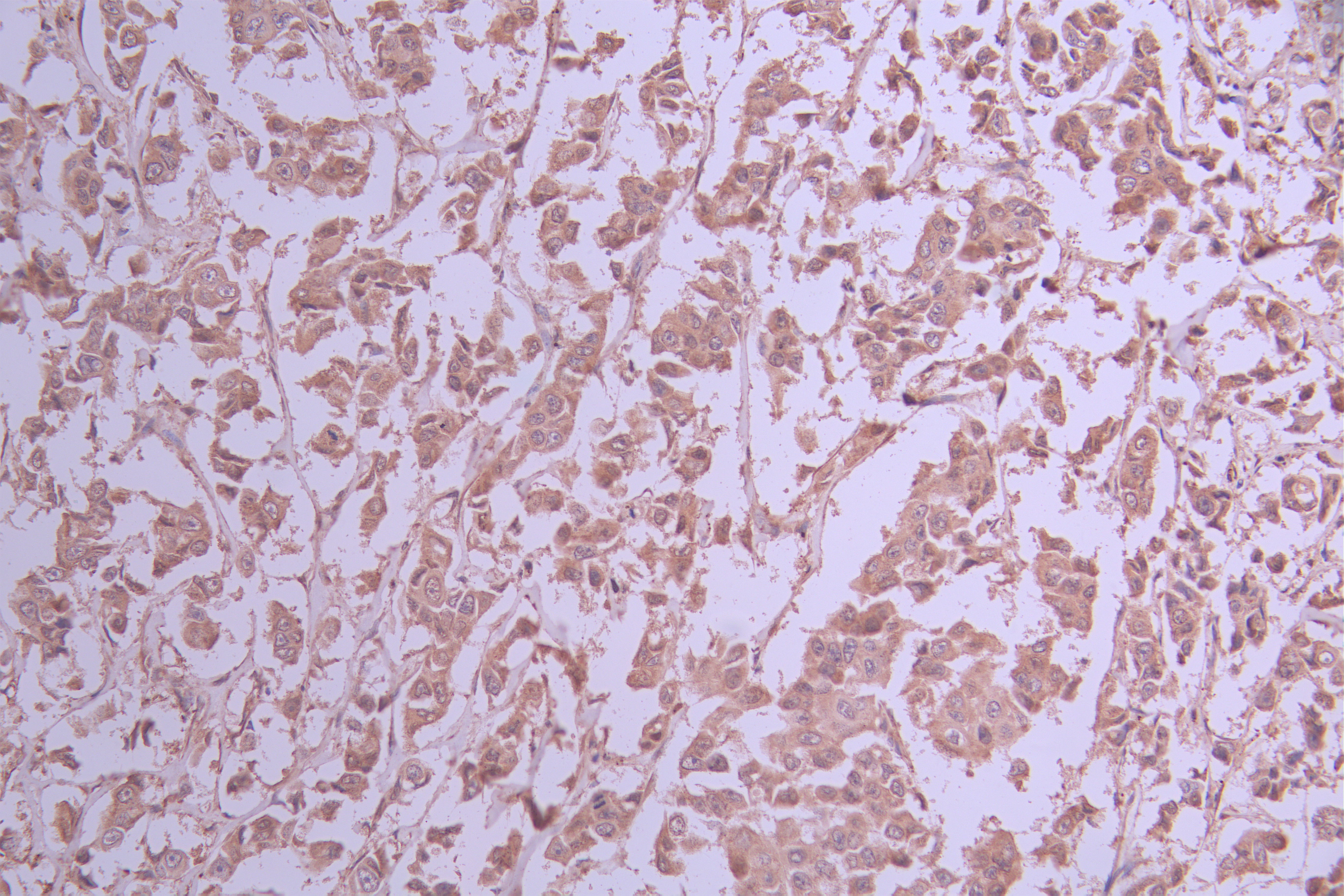
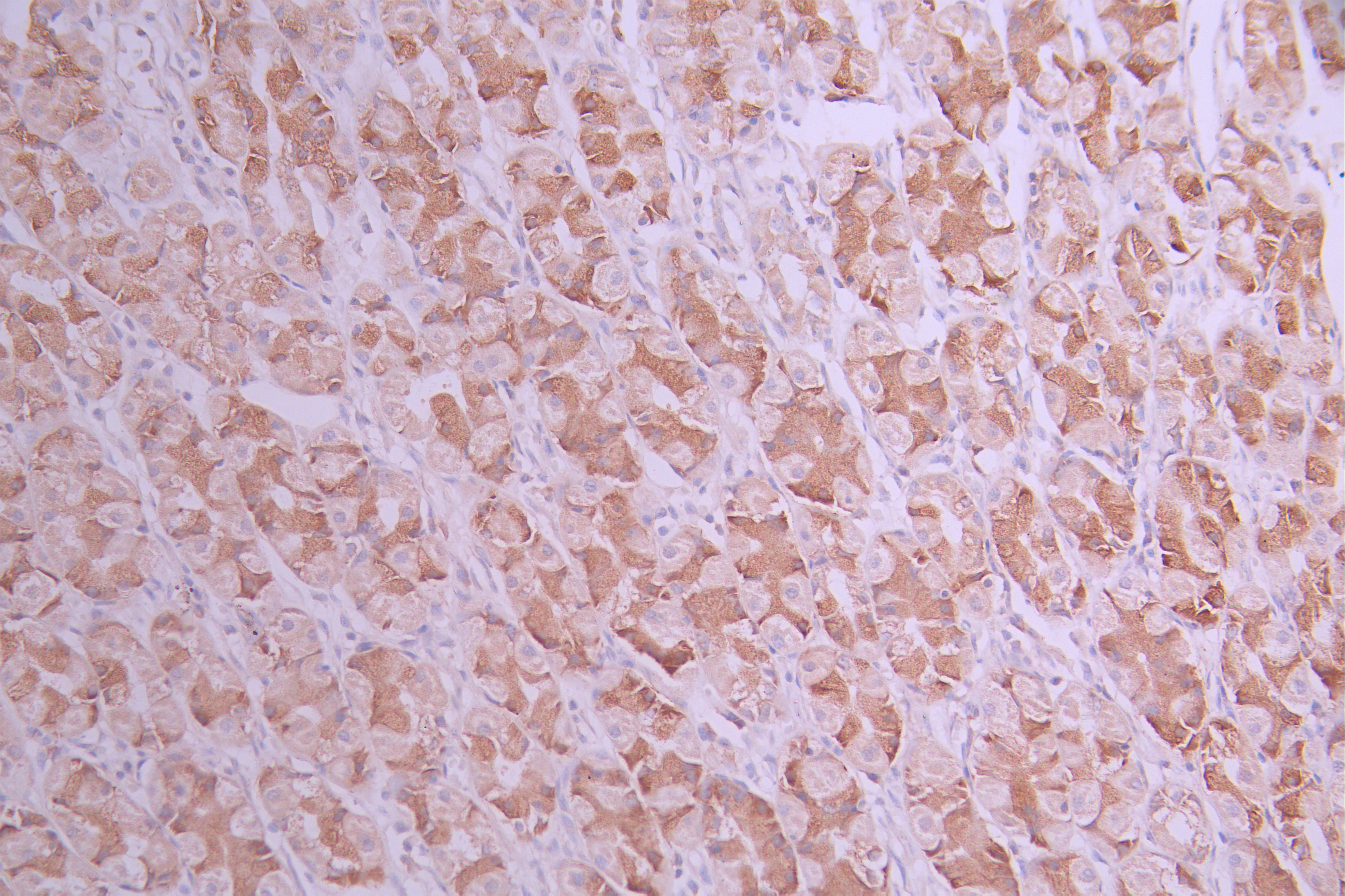
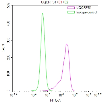
Component of the ubiquinol-cytochrome c oxidoreductase, a multisubunit transmembrane complex that is part of the mitochondrial electron transport chain which drives oxidative phosphorylation. The respiratory chain contains 3 multisubunit complexes succinate dehydrogenase (complex II, CII), ubiquinol-cytochrome c oxidoreductase (cytochrome b-c1 complex, complex III, CIII) and cytochrome c oxidase (complex IV, CIV), that cooperate to transfer electrons derived from NADH and succinate to molecular oxygen, creating an electrochemical gradient over the inner membrane that drives transmembrane transport and the ATP synthase. The cytochrome b-c1 complex catalyzes electron transfer from ubiquinol to cytochrome c, linking this redox reaction to translocation of protons across the mitochondrial inner membrane, with protons being carried across the membrane as hydrogens on the quinol. In the process called Q cycle, 2 protons are consumed from the matrix, 4 protons are released into the intermembrane space and 2 electrons are passed to cytochrome c. The Rieske protein is a catalytic core subunit containing a Component of the ubiquinol-cytochrome c oxidoreductase (cytochrome b-c1 complex, complex III, CIII). UQCRFS1 undergoes proteolytic processing once it is incorporated in the complex III dimer. One of the fragments, called subunit 9, corresponds to its mitochondrial targeting sequence (MTS). The proteolytic processing is necessary for the correct insertion of UQCRFS1 in the complex III dimer, but the persistence of UQCRFS1-derived fragments may prevent newly imported UQCRFS1 to be processed and assembled into complex III and is detrimental for the complex III structure and function.
Gene References into Functions
  1. UQCRFS1 produces N-terminal polypeptides, which remain bound to holocomplex III. We show that, in normal conditions, these UQCRFS1 fragments are rapidly removed, but when TTC19 is absent they accumulate within complex III, causing its structural and functional impairment. PMID: 28673544
  2. UQCRFS1/RISP knockdown in breast tumor cell line led to decreased mitochondrial membrane potential as well as a decrease in matrigel invasion. PMID: 21901141
  3. UQCRFS1 gene appears to be involved in development of more aggressive phenotype of breast cancer. PMID: 15047214
Subcellular Location
Mitochondrion inner membrane; Single-pass membrane protein.
Database Links

HGNC: 12587

OMIM: 191327

KEGG: hsa:7386

STRING: 9606.ENSP00000306397

UniGene: Hs.743307

icon of phone
Call us
301-363-4651 (Available 9 a.m. to 5 p.m. CST from Monday to Friday)
icon of address
Address
7505 Fannin St., Ste 610, Room 7 (CUBIO Innovation Center), Houston, TX 77054, USA
icon of social media
Join us with

Subscribe newsletter

Leave a message

* To protect against spam, please pass the CAPTCHA test below.
CAPTCHA verification
© 2007-2025 CUSABIO TECHNOLOGY LLC All rights reserved. 鄂ICP备15011166号-1
Place an order now

I. Product details

*
*
*
*

II. Contact details

*
*

III. Ship To

*
*
*
*
*
*
*

IV. Bill To

*
*
*
*
*
*
*
*