Call us
301-363-4651 (Available 9 a.m. to 5 p.m. CST from Monday to Friday)
| Code | CSB-MA000071M0m |
| Size | US$120 |
| Order now | |
| Image |
|
| Have Questions? | Leave a Message or Start an on-line Chat |
| Application | Recommended Dilution |
|---|---|
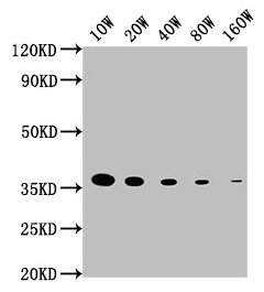
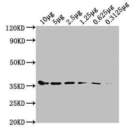
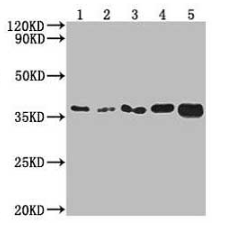
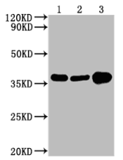
| WB | 1:5000-1:1600000 |
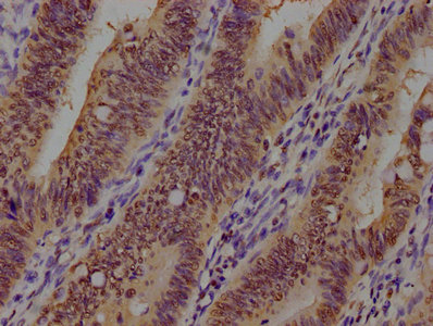
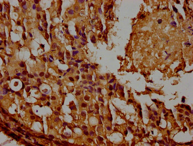
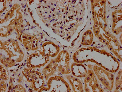
| IHC | 1:50-1:500 |
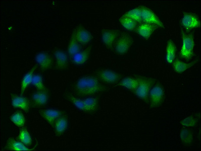
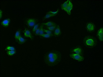
| IF | 1:50-1:200 |
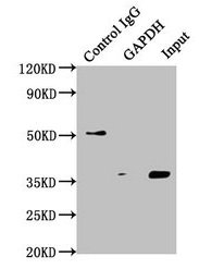
| IP | 1µl-2µl |
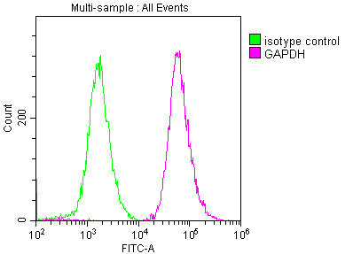
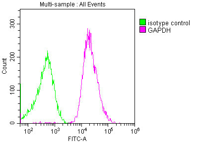
| FC | 1:100-1:300 |
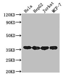
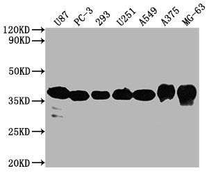
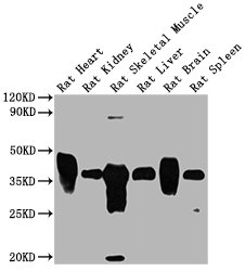
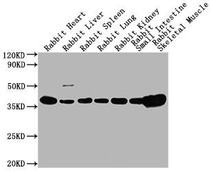
The GAPDH Monoclonal Antibody is a specific antibody that targets GAPDH. The GAPDH antibody is an internal reference antibody that functions as a loading control to ensure equal protein loading and accurate quantification of protein expression levels in different samples. This antibody can detect GAPDH in human, mouse, and rabbit species.
The immunogen used to generate this GAPDH antibody is the 2-335 amino acid region of recombinant Human GAPDH protein. The GAPDH Monoclonal Antibody is raised in mouse and belongs to the IgG1 isotype. It is purified using Protein G and reaches a purity level of greater than 95%.
The GAPDH Monoclonal Antibody is available in liquid form and has been tested in various applications, including ELISA, WB, IHC, IP, and IF. These applications make the antibody a versatile tool for the detection and analysis of GAPDH in different contexts.
Moreover, the GAPDH Monoclonal Antibody has been cited in a paper by H Miao, et al. in 2022, which highlights its utility in scientific research. The use of this validated antibody in research increases the reliability of the results and ensures reproducibility.
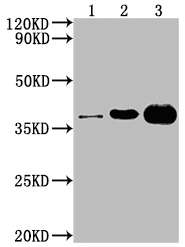
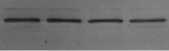
Applications : Western Blot (WB)
Sample type: Human ovarian granulosa cell carcinoma
Sample dilution: 1:1200
Review: Experiment success, antibody is usable!
By Anonymous
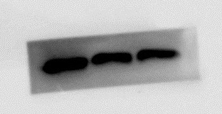
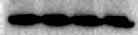
Applications : Western Blot (WB)
Sample type: KELLY
Sample dilution: 1:1000
Review: GAPDH has good results in this experiment, with correct size and single band.
By Anonymous
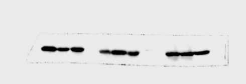
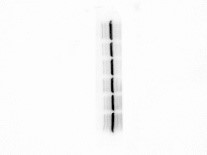
Applications : Western Blot (WB)
Sample type: Human ovarian granulosa cell carcinoma
Sample dilution: 1:1000
Review: The experiment was successful, and the antibody was successfully detected on human cells, which meets the experimental requirements and can be purchased and used in the future.
By Anonymous
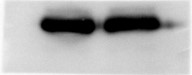
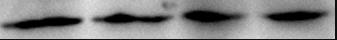
Applications : Western Blot (WB)
Sample type: Rat kidney tissue
Sample dilution: 1:5000
Review: Good antibody, good titer.
By Anonymous
Applications : Western Blot (WB)
Sample type: Mouse
Sample dilution: 1:5000
Review: Antibody specificity is very good.
By Anonymous
Applications : Western Blot (WB)
Sample type: Human 231 cell
Sample dilution: 1:2000
Review: The destination band is correct and single.
By Anonymous
Applications : Western Blot (WB)
Sample type: Cell Lysate (Chicken)
Sample dilution: 1:2000
Review: Incubate at 4 °C overnight with 1: 2000 dilution ratio, the bands are clear, the specificity is good, and it can be recycled and reused with good results.
By Anonymous
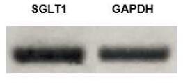
Applications : Immunoblot analyses
Review: Immunoblot analyses show the presence of SGLT-1 on BBMV of common carp.
By Anonymous
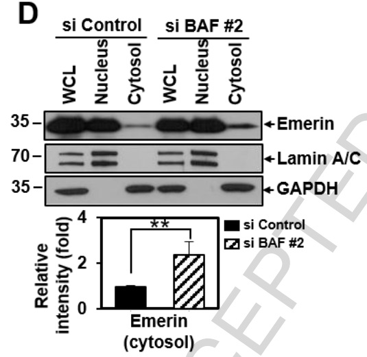
Applications : WB
Sample dilution: 1: 5000
Review: HeLa cells were treated with si BAF #2 or si Control for 72 h. Cells were fractionated and subjected to western blot analyses (upper panel). Fractionation was verified by using Lamin A/C antibody (nuclear fraction) or GAPDH antibody (cytosolic fraction). The intensity of the cytosolic emerin was analyzed using ImageJ software (lower panel).
By Anonymous
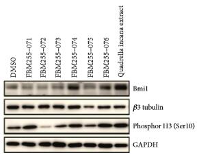
Applications : Western blot
Sample dilution: 1:1,000
Review: Western blot analysis of Bmi1, βIII tubulin, and phospho-Histone 3 (Ser 10) in NSPCs kept under proliferating media upon treatment for 3 days with DMSO, Quadrella incana leaf extract, or six different plant crude extracts. GAPDH was used as a loading control. GAPDH was used as a loading control.
By Anonymous
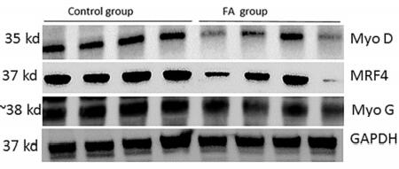
Applications : Western Blot
Sample dilution: 1:1000
Review: Protein level measurement by western blot of myogenesis regulating genes in C2C12 and GAPDH as housekeeping gene n = 4.
By Anonymous

