| Image |
-
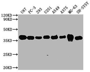
Western Blot
Positive WB detected in: U87 whole cell lysate, PC3 whole cell lysate, 293 whole cell lysate, U251 whole cell lysate, A549 whole cell lysate, MG-63 whole cell lysate, U251 whole cell lysate, SH-SY5Y whole cell lysate
All lanes GAPDH antibody at 1:5000
Secondary
Goat polyclonal to mouse IgG at 1/50000 dilution
Predicted band size: 36 KDa
Observed band size: 36 KDa
Exposure time: 5min
-
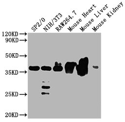
Western Blot
Positive WB detected in: SP2/0 whole cell lysate, NIH/3T3 whole cell lysate, Raw264.7 whole cell lysate, Mouse heart tissue, Mouse kidney tissue
All lanes GAPDH antibody at 1:5000
Secondary
Goat polyclonal to mouse IgG at 1/50000 dilution
Predicted band size: 36 KDa
Observed band size: 36 KDa
Exposure time: 5min
-
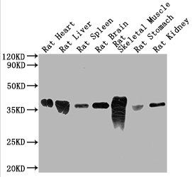
Western Blot
Positive WB detected in: Rat heart tissue, Rat liver tissue, Rat spleen tissue, Rat brain tissue, Rat skeletal tissue, Rat stomach tissue, Rat kidney tissue
All lanes GAPDH antibody at 1:5000
Secondary
Goat polyclonal to mouse IgG at 1/50000 dilution
Predicted band size: 36 KDa
Observed band size: 36 KDa
Exposure time: 5min
-
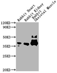
Western Blot
Positive WB detected in: Rabbit heart tissue, Rabbit kidney tissue, Rabbit skeletal muscle tissue
All lanes GAPDH antibody at 1:5000
Secondary
Goat polyclonal to mouse IgG at 1/50000 dilution
Predicted band size: 36 KDa
Observed band size: 36 KDa
Exposure time: 5min
-
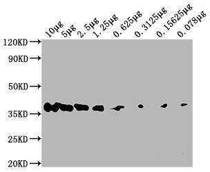
Western Blot
Positive WB detected in: Hela whole cell lysate at 10μg, 5μg, 2.5μg, 1.25μg, 0.625μg, 0.3125μg, 0.15625μg, 0.078μg All lanes:GAPDH antibody at 1:5000
Secondary
Goat polyclonal to mouse IgG at 1/50000 dilution
Predicted band size: 36 KDa
Observed band size: 36 KDa
Exposure time: 5min
-
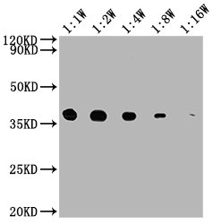
Western Blot
Positive WB detected in: 15μg hela whole cell lysate GAPDH antibody at 1:10000, 1:20000, 1:40000, 1:80000, 1:160000
Secondary
Goat polyclonal to mouse IgG at 1/50000 dilution
Predicted band size: 36 KDa
Observed band size: 36 KDa
Exposure time: 5min
-
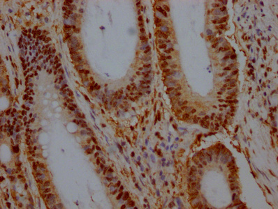
IHC image of CSB-MA000071M2m diluted at 1:500 and staining in paraffin-embedded human colon cancer performed on a Leica BondTM system. After dewaxing and hydration, antigen retrieval was mediated by high pressure in a citrate buffer (pH 6.0). Section was blocked with 10% normal goat serum 30min at RT. Then primary antibody (1% BSA) was incubated at 4°C overnight. The primary is detected by a Goat anti-rabbit polymer IgG labeled by HRP and visualized using 0.05% DAB.
-
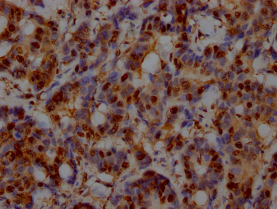
IHC image of CSB-MA000071M2m diluted at 1:500 and staining in paraffin-embedded human breast cancer performed on a Leica BondTM system. After dewaxing and hydration, antigen retrieval was mediated by high pressure in a citrate buffer (pH 6.0). Section was blocked with 10% normal goat serum 30min at RT. Then primary antibody (1% BSA) was incubated at 4°C overnight. The primary is detected by a Goat anti-rabbit polymer IgG labeled by HRP and visualized using 0.05% DAB.
-
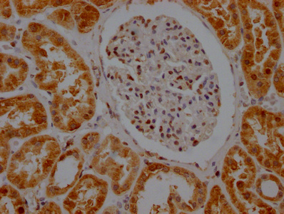
IHC image of CSB-MA000071M2m diluted at 1:500 and staining in paraffin-embedded human kidney tissue performed on a Leica BondTM system. After dewaxing and hydration, antigen retrieval was mediated by high pressure in a citrate buffer (pH 6.0). Section was blocked with 10% normal goat serum 30min at RT. Then primary antibody (1% BSA) was incubated at 4°C overnight. The primary is detected by a Goat anti-rabbit polymer IgG labeled by HRP and visualized using 0.05% DAB.
-
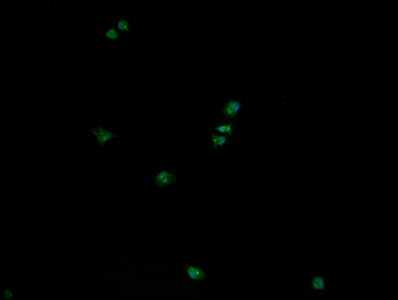
Immunofluorescence staining of Hela cells with (CSB-MA000071M2m)at 1:50, counter-stained with DAPI. The cells were fixed in 4% formaldehyde, permeabilized using 0.2% Triton X-100 and blocked in 10% normal Goat Serum. The cells were then incubated with the antibody overnight at 4°C. Nuclear DNA was labeled in blue with DAPI. The secondary antibody was FITC-conjugated AffiniPure Goat Anti-Mouse IgG (H+L).
-
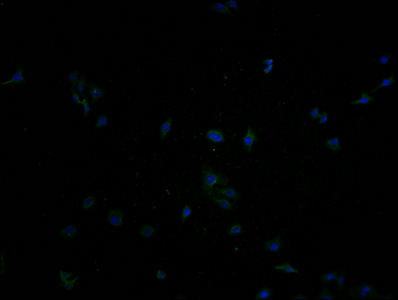
Immunofluorescence staining of HepG2 cells with(CSB-MA000071M2m)at 1:50, counter-stained with DAPI. The cells were fixed in 4% formaldehyde, permeabilized using 0.2% Triton X-100 and blocked in 10% normal Goat Serum. The cells were then incubated with the antibody overnight at 4°C. Nuclear DNA was labeled in blue with DAPI. The secondary antibody was FITC-conjugated AffiniPure Goat Anti-Mouse IgG (H+L).
-
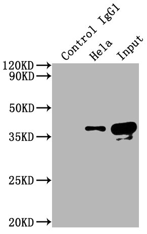
Immunoprecipitating GAPDH in Hela whole cell lysate
Lane 1: Mouse control IgG instead of CSB-MA000071M2m in Hela whole cell lysate.
Lane 2: CSB-MA000071M2m (5µl) + Hela whole cell lysate (500µg)
Lane 3: Hela whole cell lysate (10µg)
For western blotting, the blot was detected with CSB-MA000071M2m at 1:5000, and a HRP-conjugated Protein G antibody was used as the secondary antibody at 1:2000
|