Call us
301-363-4651 (Available 9 a.m. to 5 p.m. CST from Monday to Friday)
| Code | CSB-MA000071M1m |
| Size | US$120 |
| Order now | |
| Image |
|
| Have Questions? | Leave a Message or Start an on-line Chat |
| Application | Recommended Dilution |
|---|---|
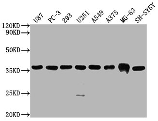
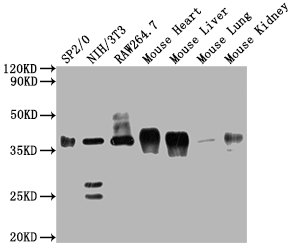
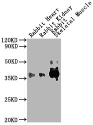
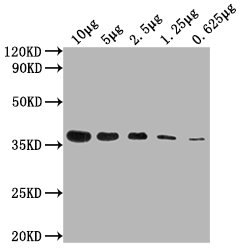
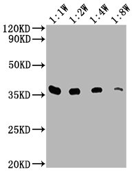
| WB | 1:5000-1:80000 |
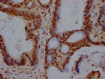
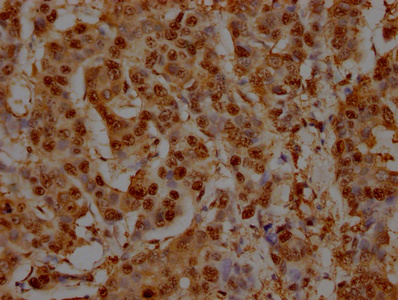
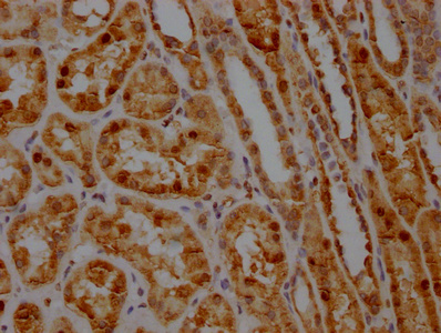
| IHC | 1:200-1:500 |
| IF | 1:50-1:100 |
| IP | 2µl-8µl |
This GAPDH monoclonal antibody was raised by fusion of B lymphocytes with immortal cell cultures to produce hybridomas (A Recombinant Human GAPDH protein was used in the immunization process). Hybridomas would produce many copies of GAPDH monoclonal antibody. The specificity of this GAPDH monoclonal antibody makes it extremely efficient for binding of antigen within a mixture of GAPDH. In addition, this antibody has been validated in ELISA, WB, IHC, IP, IF.
GAPDH (G3PD) is the abbreviation of glyceraldehyde-3-phosphate dehydrogenase, which is an enzyme in glycolysis and consists of 4 subunits of 30-40 kDa. The molecular weight is 146 kDa. The enzyme gene is a house-keeping gene, which is expressed at a high level in almost all tissues. The protein expression level in the same cell or tissue is generally constant and is not induced by the partial recognition sites contained. The influence of the substance remains constant, so it is widely used as a standardized internal reference for the extraction of total RNA, poly(A)+ RNA, Western blot and other experimental operations.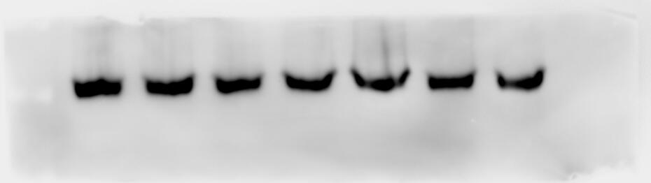
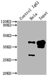
Applications : Western Blot (WB)
Sample type: hek293 cell
Sample dilution: 1:5000
Review: 使用购买的二抗稀释液1:5000稀释此产品,4℃孵育过夜,效果较好,甚至比以前用过的1:1000的GAPDH抗体条带更加整齐明亮 The product was diluted 1:5000 with the purchased secondary antibody dilution and incubated overnight at 4°C. The results were better and even brighter than the previously used 1:1000 GAPDH antibody bands.
By Anonymous
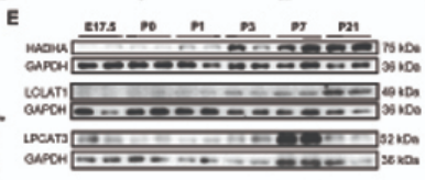
Applications : Immunoblot analyses
Sample type: cells
Review: Immunoblot analyses of protein levels of Hadha, Lclat1, and Lpcat3 across cardiac development. Images presented were representative of 2 experiments. Relative levels of individual proteins were normalized to that of GAPDH, and illustrated as bar plots on the lower panel.
By Anonymous
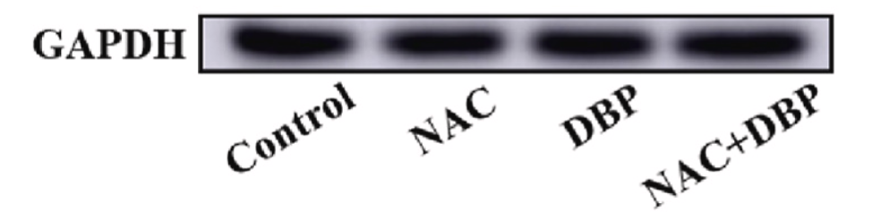
Applications : Western blot
Sample type: cells
Review: GAPDH was served as protein loading controls.
By Anonymous
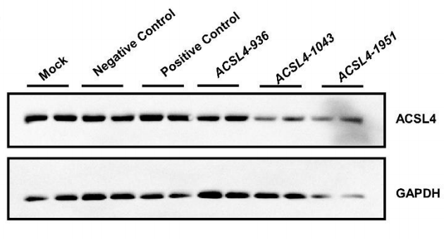
Applications : Western blot(WB)
Sample type: cells
Review: Western blot for identification of ACSL4 knockdown efficiency. Three ACSL4 siRNA lines (ACSL4-936, ACSL4-1043, and ACSL4-1951) were tested (n = 2).
By Anonymous
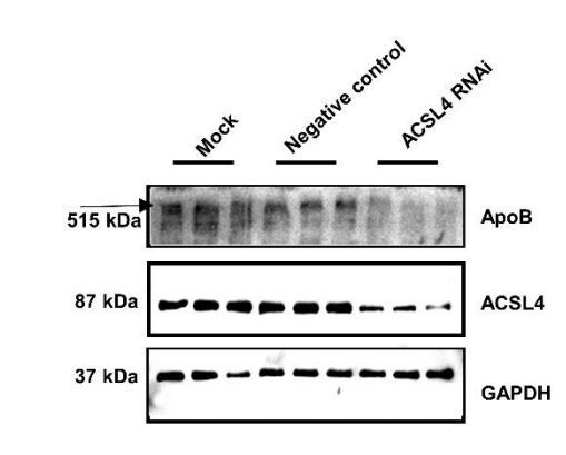
Applications : Western blot(WB)
Sample type: cells
Review: Western blot analysis of ACSL4 and ApoB expression levels in HepG2 cells. (n = 3).
By Anonymous

