
Call us
301-363-4651 (Available 9 a.m. to 5 p.m. CST from Monday to Friday)

| Code | CSB-MA000051M0m |
| Size | US$120 |
| Order now | |
| Image |
|
| Have Questions? | Leave a Message or Start an on-line Chat |
| Application | Recommended Dilution |
|---|---|
| WB | 1:500-1:5000 |
| IP | 1:200-1:2000 |
Green Fluorescent Protein (GFP) is a highly versatile and widely adopted genetic tag in molecular and cellular biology. As a self-sufficient fluorescent marker, it enables researchers to visually track gene expression, monitor protein localization in live cells, and investigate dynamic cellular processes in real-time without the need for external substrates. Its compact size and minimal impact on the native conformation of target proteins make it an invaluable asset for functional protein studies. This mouse GFP monoclonal antibody (IgG2b isotype) is produced by immunizing mice with recombinant GFP protein and subsequently purified via Protein G affinity chromatography, ensuring a high purity level of over 95%. It demonstrates exceptional specificity and reliability across multiple assay platforms.
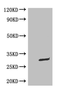
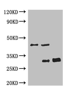
Rigorous in-house validation confirms its efficacy in Western Blot (WB), Enzyme-Linked Immunosorbent Assay (ELISA), and Immunoprecipitation (IP) applications. In WB experiments using transfected cell lysates, the antibody detects the GFP-tagged fusion protein with a precisely observed molecular weight of 30 kD, closely matching the theoretical prediction. Furthermore, its robust performance in IP workflows effectively validates its capability to specifically precipitate GFP-tagged targets from complex biological samples, making it an excellent reagent for studying protein-protein interactions.
As a rigorously validated immunological tool, this antibody serves as a critical reagent for confirming recombinant protein expression, facilitating affinity purification, and supporting a wide range of downstream biochemical analyses. With a proven track record of citation in peer-reviewed scientific literature (3 citations), this CUSABIO monoclonal antibody is backed by comprehensive technical support, ensuring dependable and reproducible results to accelerate your cutting-edge research.
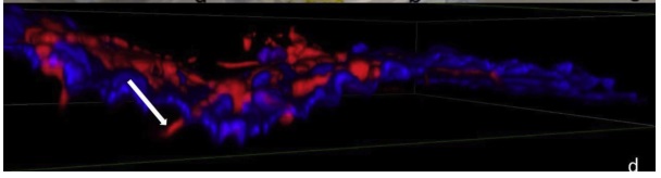
Applications : IHC
Sample dilution: 1: 500
Review: fluorescent microscopic analysis 12 h after inoculation showing the fungal hyphae expressing GFP (in red) penetrating the floral tissue (in blue).
By Anonymous
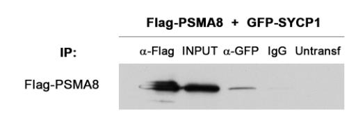
Applications : IP
Sample dilution: 1: 1000
Review: HEK293T cells were transfected with Flag-PSMA8 and GFP-SYCP1. Protein complexes were immunoprecipitated overnight with either an anti-Flag or anti-EGFP or IgGs (negative control), and were analyzed by immunoblotting with the indicated antibody. PSMA8 co-immunoprecipitates with SYCP1.
By Anonymous
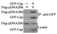
Applications : IP
Review: PCV2 Cap interacts with pDNAJB6 in transfected cells. HEK293T cells were co-transfected with GFP-Cap and Flag-pDNAJB6 expression plasmids or co-transfected with the pEGFP-N1 control vector and the Flag-pDNAJB6 expression plasmid. The cells transfected with pEGFP-N1 control vector, GFP-Cap expression plasmid or Flag-pDNAJB6 expression plasmid alone served as controls. The interaction of GFP-Cap with Flag-pDNAJB6 was identified through immunoprecipitation using anti-Flag antibodies or anti-GFP antibodies.
By Anonymous