| Image |
-
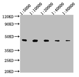
Western Blot
Positive WB detected in: 50ng V5-tagged fusion protein
V5 Tag antibody at 1:5000, 1:10000, 1:20000, 1:40000, 1:80000
Secondary
Goat polyclonal to mouse IgG at 1/50000 dilution
Predicted band size: 37.8 KDa
Observed band size: 43 KDa
Exposure time: 10S
-
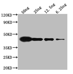
Western Blot
Positive WB detected in: Mouse anti V5 tag Monoclonal antibody at 1:5000
Lane 1: Recombinant V5-tagged fusion protein at 50ng
Lane 2: Recombinant V5-tagged fusion protein at 25ng
Lane 3: Recombinant V5-tagged fusion protein at 12.25ng
Lane 4: Recombinant V5-tagged fusion protein at 6.25ng
Secondary
Goat polyclonal to Mouse IgG at 1/50000 dilution
Predicted band size: 37.8 kd
Observed band size: 43 kd
Exposure time: 5min
-
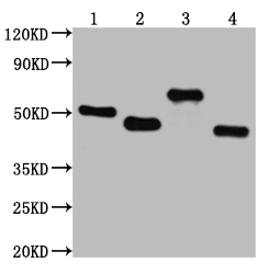
Western Blot
Positive WB detected in: V5-tagged fusion protein 1, 2, 3, 4 at 50ng
All lanes: V5 antibody at 1:2000
Secondary
Goat polyclonal to mouse IgG at 1/50000 dilution
Predicted band size: 50.2, 47.6, 66.7, 37.8 KDa
Observed band size: 52, 47, 67, 42 KDa
Exposure time: 30s
-
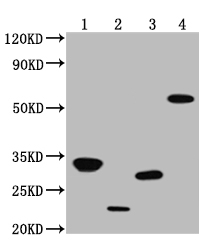
Western Blot
Positive WB detected in: V5-tagged fusion protein 1, 2, 3, 4, at 50ng
All lanes: V5 antibody at 1:2000
Secondary
Goat polyclonal to mouse IgG at 1/50000 dilution
Predicted band size: 31.9, 20.4, 24.2, 53.4 KDa
Observed band size: 32, 21, 28, 58 KDa
Exposure time: 5min
-
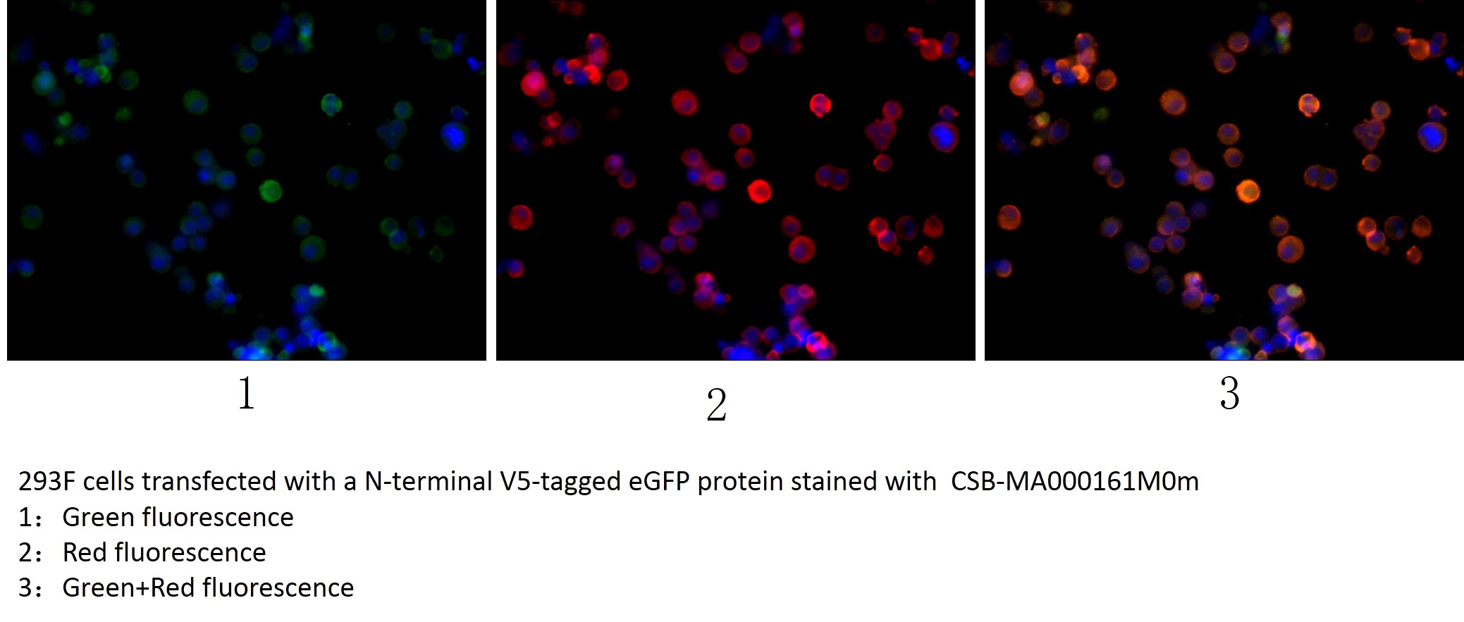
Immunofluorescence staining of transfected HEK293F cells with CSB-MA000161M0m at 1:100, counter-stained with DAPI. The cells were fixed in 4% formaldehyde, permeabilized using 0.2% Triton X-100 and blocked in 10% normal Goat Serum. The cells were then incubated with the antibody overnight at 4°C. The secondary antibody was Alexa Fluor R-PE Goat Anti-Mouse IgG(H+L).
-
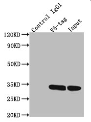
Immunoprecipitating V5 Tag in Transfected HEK293F cells whole cell lysate
Lane 1: Mouse control IgG1 instead of CSB-MA000161M0m in Transfected HEK293F cells whole cell lysate
Lane 2: CSB-MA000161M0m (2.5µl) + Transfected HEK293F cells whole cell lysate (500µg)
Lane 3: Transfected HEK293F cells whole cell lysate (20µg)
For western blotting, the blot was detected with CSB-MA000161M0m at 1:5000, and a HRP-conjugated Protein G antibody was used as the secondary antibody at 1:50000
-
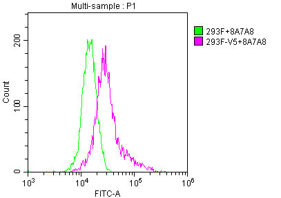
Overlay histogram showing untransfected HEK293F cells (green line) or transfected HEK293F cells (red line) stained with CSB-MA000161M0m. The cells were fixed with 70% Ethylalcohol (18h) and then permeabilized with 0.3% Triton X-100 for 2 min. The cells were then incubated in 10% normal goat serum to block non-specific protein-protein interactions followed by the antibody (1:200/1*106cells) for 1 h at 4°C. The secondary antibody used was FITC goat anti-mouse IgG(H+L) at 1/100 dilution for 30min at 4°C. Acquisition of >10,000 events was performed.
|