Call us
301-363-4651 (Available 9 a.m. to 5 p.m. CST from Monday to Friday)
ACVR1C/ALK7, as a key member of the TGF-β superfamily, has recently garnered significant attention due to its dual roles in metabolic regulation and tumor progression. It not only plays crucial roles in adipose tissue differentiation, insulin sensitivity, and energy balance but also exhibits "context-dependent" functions in various solid tumors, either inhibiting or promoting tumor growth. This article systematically reviews the molecular mechanisms, signaling pathways, and functions of ACVR1C/ALK7 in metabolic diseases and cancer, aiming to assist your related research.
1. Research Background and Biomedical Significance of ACVR1C/ALK7
2. Molecular Characteristics, Structural Basis, and Ligand Recognition of ACVR1C/ALK7
3. ACVR1C/ALK7-Mediated Signal Transduction Pathways
4. The Role of ACVR1C/ALK7 in Disease Pathogenesis and Progression
ACVR1C (Activin receptor-like kinase 7, ALK7) is a type I receptor member of the TGF-β superfamily, possessing a typical receptor serine/threonine kinase domain and a GS regulatory region. Its function relies on forming complexes with type II receptors to mediate downstream signaling cascades. Early cloning and expression profiling studies revealed that ACVR1C exhibits significant spatiotemporal specificity in embryonic development and adult tissues, providing foundational evidence for its roles in developmental regulation and adult tissue homeostasis. Although its kinase core is highly conserved with ALK4/ALK5, minor differences in the extracellular domain significantly affect ligand affinity and signal output strength, endowing ACVR1C with functional uniqueness.
At the signal transduction level, ACVR1C primarily activates the canonical TGF-β-like pathway mediated by Smad2/3, while concurrently triggering non-canonical pathways such as MAPK and PI3K/AKT under specific cellular conditions, thereby achieving integrated regulation of cell proliferation, differentiation, migration, and metabolism. Growing evidence indicates that its signal output highly depends on ligand type, receptor complex composition, and intracellular regulatory factors (such as I-Smads and E3 ubiquitin ligases), providing a mechanistic basis for explaining the opposing effects of ACVR1C in different physiological and pathological contexts. However, current research predominantly relies on in vitro overexpression systems or broad-spectrum inhibitors, limiting the precise elucidation of ACVR1C's intrinsic functions and its interplay with other type I receptors, highlighting the necessity for developing selective tools and conditional in vivo models.
Functional studies have closely linked ACVR1C to embryonic development, adipose tissue homeostasis, and energy metabolism. Gene knockout and developmental models suggest its involvement in stem cell fate determination and organogenesis. In adult metabolic tissues, ACVR1C influences insulin sensitivity and energy balance by inhibiting preadipocyte proliferation and promoting differentiation, making it a focal point in obesity and type 2 diabetes research. Conversely, in cancer and fibrosis studies, the role of ACVR1C exhibits a clear duality: it can exert inhibitory effects in some tumor contexts while promoting invasion and migration in specific microenvironments, reflecting the decisive influence of cellular context and ligand repertoire on its signal output. Due to limited human genetic and clinical association evidence, establishing a causal relationship between ACVR1C and disease phenotypes at the genetic level remains a major challenge in the field.
ACVR1C belongs to the TGF-β superfamily type I receptors, comprising an extracellular ligand-binding domain, a single transmembrane helix, and intracellular GS and kinase domains. It was initially cloned as an orphan receptor and later confirmed as a type I receptor for Nodal and Activin-like ligands [1]. Structural and binding studies indicate that Nodal forms a multimolecular complex with Cripto and ALK4/ALK7, where key amino acid residues determine ligand selectivity [2]. Notably, Activin-like ligands exhibit dual specificity: primarily activating Smad2/3 via ALK4/ALK7, but some dimers can also activate Smad1/5 via ALK2, suggesting that ligand composition and receptor pairing jointly determine signaling branch selection [3].
Transcriptomic and molecular studies consistently show that ACVR1C is highly enriched in white and brown adipose tissues and significantly upregulated during the late stages of adipocyte differentiation [4]. Human adipose tissue studies further confirm that its expression is negatively correlated with obesity and closely related to glycolipid metabolism indicators, supporting the role of the Activin B–ALK7 axis in adipose tissue homeostasis regulation [5]. Furthermore, ACVR1C is also expressed in the nervous system and pancreatic β-cells, with various splice variants exhibiting differences in signaling capacity [6]. For example, Alk7-v3 cannot effectively transduce Nodal-Cripto signals, suggesting that transcriptional variants can reshape signal output without altering receptor expression. Its expression can also be regulated by metabolic stimuli; for instance, β3-adrenergic agonists downregulate ALK7 expression in brown adipose tissue, reflecting its cross-regulation with sympathetic-metabolic pathways [6]. Recent transcriptomic studies have also linked ACVR1C to neuroendocrine development, showing that it inhibits Mkrn3 expression via Smad2/3-mediated chromatin regulation, thereby affecting puberty onset [7].
At the ligand level, Activin E (INHBE) is considered a crucial functional ligand for ACVR1C, activating the Smad2/3 pathway via this receptor and regulating energy metabolism-related gene expression [9][10]. Pharmacological blockade experiments show that the small-molecule inhibitor SB431542 inhibits Activin E-induced Smad2/3 phosphorylation and downstream transcriptional activation, supporting its signaling through ALK7 [10]. Embryonic model studies indicate that Nodal signaling primarily relies on ALK4 in early developmental stages, while ALK7 is not essential for embryogenesis and is more likely to perform tissue-specific or ligand-specific functions [11-13].
Functionally, the role of the Activin E–ACVR1C axis in metabolic regulation exhibits phenotypic disparities. On one hand, mouse in vivo studies suggest this pathway inhibits lipolysis, promotes fat storage, and suppresses PPARγ-related genes, potentially increasing metabolic risk [9]; on the other hand, in vitro studies on brown/beige adipocytes show Activin E can upregulate Ucp1 and Fgf21 via ALK7/Smad2/3, indicating its role in promoting thermogenesis and energy expenditure in specific lineages [10]. These discrepancies can be explained by differences in research models, cell types, dosages, timing, and receptor microenvironment, emphasizing the critical roles of signal bias and chromatin context in determining transcriptional outcomes. Due to the lack of systematic affinity measurements and high-resolution structural data, the molecular basis of ligand-receptor selectivity remains to be elucidated [11].
ACVR1C/ALK7, as a key node in the TGF-β superfamily signaling network, converts extracellular ligand stimuli into multi-layered intracellular responses. Its signal output includes both the classic transcriptional regulatory axis centered on Smad2/3 and various non-canonical signaling branches. The division of labor among different pathways in terms of timescale, spatial localization, and biological effects results in significant context-dependent functionality for ACVR1C.
In the classic model, ligand binding promotes the formation of a complex between the type II receptor and ACVR1C. The type II receptor phosphorylates the GS region of ACVR1C, thereby activating its kinase activity and recruiting Smad2/3. Phosphorylated Smad2/3 forms a complex with co-Smad4, translocates into the nucleus, and regulates target gene transcription [14]. This axis plays a central role in adipocyte differentiation, pancreatic β-cell function, and fate determination in various tumor cells.
In tumor models, ACVR1C-Smad2 signaling has been shown to promote tumor cell invasion and growth in specific contexts. For example, in retinoblastoma, upregulated ACVR1C expression accompanies enhanced Smad2 activity, and inhibiting this pathway significantly reduces invasion and survival capacity, while the role of Smad3 is relatively limited [16]. These results suggest that despite high structural similarity, Smad2 and Smad3 may bear different functional weights in ACVR1C signal output, with differences potentially determined by receptor-ligand combinations and nuclear co-factors [15].
Besides the canonical Smad pathway, ACVR1C can also mediate non-canonical signals via β-arrestin, Ral GTPase, etc. Related studies show that Nodal-ALK7 signaling can form stable complexes in the cytoplasm, regulating receptor internalization, ERK1/2 activation, and cytoskeleton remodeling, thereby rapidly affecting cell migration and invasion behavior [17]. In pancreatic cancer models, ALK7 promotes basement membrane disruption and tumor cell intravasation via the β-catenin/MMP axis, emphasizing the role of its non-Smad branches in the metastasis process [18].
Furthermore, ACVR1C can regulate macrophage and vascular smooth muscle cell phenotypes by inhibiting PPARγ expression, promoting inflammatory responses and vascular remodeling [19][20]. These findings indicate that ACVR1C's signal output is not a single pathway but is shaped by multiple parallel pathways, with bias depending on cell type, ligand concentration, and receptor microenvironment.
In various solid tumors, ACVR1C can induce cell apoptosis and inhibit tumor dissemination via Smad2/3-mediated transcriptional programs, regarded as an intrinsic tissue homeostasis barrier [23],[24]. Clinical sample analyses show that reduced ACVR1C expression is often associated with poor prognosis, such as in gallbladder cancer and breast cancer [25-28].
However, in contexts like colorectal cancer, retinoblastoma, and pancreatic ductal adenocarcinoma, ACVR1C can be repurposed by tumor cells to promote EMT, stemness maintenance, and metastasis through autocrine or paracrine loops [29-32]. These contradictory phenomena highlight the highly context-dependent nature of ACVR1C in cancer, with its function contingent upon the ligand repertoire, signaling pathway bias, and tumor microenvironment state.
Population genetics studies provide strong evidence for ACVR1C's causal role in human body fat distribution. Rare variant analysis from the UK Biobank shows that loss-of-function of ACVR1C is associated with more favorable fat distribution and lipid profiles [38]. Functional studies further indicate that ACVR1C affects fat storage and insulin sensitivity by inhibiting lipolysis and regulating PPARγ signaling [21].
It is important to note that ACVR1C is also involved in regulating insulin gene expression and cell survival in pancreatic β-cells, and excessive inhibition of this pathway may pose risks to islet function [42-44]. Therefore, its development as a therapeutic target for metabolic diseases requires emphasis on tissue specificity and safety window evaluation.
In the cardiovascular system, ACVR1C can inhibit pathological cardiac hypertrophy under pressure overload conditions, while its activation in a diabetic context may promote myocardial apoptosis and fibrosis [45-47]. In pregnancy-related diseases, Nodal-ALK7 signaling inhibits trophoblast cell invasion and is believed to be associated with preeclampsia occurrence [48][49].
In the nervous system, ACVR1C is involved in post-ischemic neuroprotection and also plays a role in synaptic plasticity and memory formation [8], suggesting its potential for different intervention values in acute injury versus chronic neurodegenerative changes.
ACVR1C/ALK7, as an important signaling receptor in the TGF-β superfamily, plays key roles in various physiological and pathological processes such as metabolic regulation and tumor progression, and has recently become an emerging therapeutic target. Current drug development targeting ACVR1C mainly focuses on different strategies including siRNA, small-molecule inhibitors, and monoclonal antibodies, covering diseases such as obesity, cancer, diabetic nephropathy, and SARS-CoV-2 infection. Some pipelines under investigation are summarized in the table below:
| Drug | Mechanism of Action | Drug Type | Indication Under Investigation | Research Institution | Highest R&D Phase |
|---|---|---|---|---|---|
| ARO-ALK7 | ALK7 Inhibitor | siRNA | Obesity | Arrowhead Pharmaceuticals, Inc. | Clinical I/II |
| A-83-01 | ALK4/5/7 Inhibitor | Small Molecule | Cancer | Japanese Foundation for Cancer Research | Preclinical |
| ALN-2232 | ALK7 Inhibitor | siRNA | Obesity | Alnylam Pharmaceuticals, Inc. | Preclinical |
| SB-431542 | ALK4/5/7 Inhibitor | Small Molecule | SARS-CoV-2 Infection | Indian Council of Medical Research | Preclinical |
| ALK7 siRNA (Shian Bio) | ALK7 Inhibitor | siRNA | Obesity | Suzhou Shian Biotechnology Co., Ltd. | Preclinical |
| ITI-8000 | ALK7 Inhibitor | mAb (Monoclonal Antibody) | Cancer | Immunomic Therapeutics, Inc. | Preclinical |
| Protocatechuic Aldehyde | ALK7 Modulator | Small Molecule | Diabetic Ulcer | Shuguang Hospital Affiliated to Shanghai University of Traditional Chinese Medicine | Preclinical |
| SB-505124 | ALK4/5/7 Inhibitor | Small Molecule | \- | GSK Plc | Preclinical |
| SGB-ALK7 | ALK7 Inhibitor | siRNA | Obesity | Suzhou SGB Pharmaceuticals Co., Ltd. | Preclinical |
(Data as of December 26, 2025, sourced from Synapse)
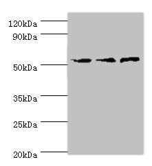
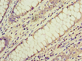
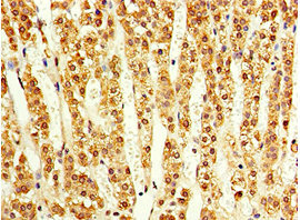
ACVR1C/ALK7 is a crucial signaling node connecting developmental biology, metabolic regulation, and disease pathology. Its biological effects highly depend on cell type, ligand environment, and signal bias, which endows it with therapeutic potential but also increases translational difficulty. CUSABIO provides ACVR1C/ALK7 antibody products to support your related mechanism research and targeted drug development.
References
[1] Masahiro Kogame, Shinji Matsuo, Masashi Nakatani, Akira Kurisaki, Hiromu Nishitani, Kunihiro Tsuchida, Hiromu Sugino.(2006). ALK7 is a novel marker for adipocyte differentiation.
[2] Luisa Calvanese, Annamaria Sandomenico, Andrea Caporale, Annalia Focà, Giuseppina Focà, Gabriella D'Auria, Lucia Falcigno, Menotti Ruvo.(2015). Conformational features and binding affinities to Cripto, ALK7 and ALK4 of Nodal synthetic fragments.
[3] Oddrun Elise Olsen, Hanne Hella, Samah Elsaadi, Carsten Jacobi, Erik Martı́nez-Hackert, Toril Holien.(2020). Activins as Dual Specificity TGF-β Family Molecules: SMAD-Activation via Activin- and BMP-Type 1 Receptors.
[4] Yan Song, Jinsoo Ahn, Yeunsu Suh, Michael E Davis, Kichoon Lee.(2013). Identification of novel tissue-specific genes by analysis of microarray databases: a human and mouse model.
[5] Lena M S Carlsson, Peter Jacobson, Andrew Walley, Philippe Froguel, Lars Sjöström, Per-Arne Svensson, Kajsa Sjöholm.(2009). ALK7 expression is specific for adipose tissue, reduced in obesity and correlates to factors implicated in metabolic disease.
[6] Masaru Murakami, Mitsuyuki Shirai, Ryo Ooishi, Asako Tsuburaya, Kumiko Asai, Osamu Hashimoto, Kenji Ogawa, Yoshii Nishino, Masayuki Funaba.(2013). Expression of activin receptor-like kinase 7 in adipose tissues.
[7] Dor Shalev, G. Golan, Lilach Pnueli, Anat Kahan, Yael Mandel‐Gutfreund, Philippa Melamed.(2025). High-throughput transcriptome analysis reveals a developmental increase in Acvr1c which mediates epigenetic repression of the gene encoding the pubertal brake, Makorin ring finger protein 3.
[8] Meilian Liu, Yudie Li, Song Han, Hongyu Wang, Junfa Li.(2022). Activin A alleviates neuronal injury through inhibiting cGAS-STING-mediated autophagy in mice with ischemic stroke.
[9] Rene C. Adam, Dwaine S. Pryce, Joseph S. Lee, Yuanqi Zhao, Ivory J. Mintah, Soo Min, Gábor Halász, Jason Mastaitis, Gurinder S. Atwal, Senem Aykul, Vincent Idone, Aris N. Economides, Luca A. Lotta, Andrew Murphy, George D. Yancopoulos, Mark W. Sleeman, Viktoria Gusarova.(2023). Activin E–ACVR1C cross talk controls energy storage via suppression of adipose lipolysis in mice.
[10] Maho Sakaki, Yuji Kamatari, Akira Kurisaki, Masayuki Funaba, Osamu Hashimoto.(2024). Activin E upregulates uncoupling protein 1 and fibroblast growth factor 21 in brown adipocytes.
[11] .(2021). Decision letter: Regulation of Nodal signaling propagation by receptor interactions and positive feedback.
[12] Hannes Preiß, Anna C. Kögler, David Mörsdorf, Daniel Čapek, Gary H. Soh, Katherine W. Rogers, Hernán Morales‐Navarrete, María Almuedo‐Castillo, Patrick Müller.(2022). Author response: Regulation of Nodal signaling propagation by receptor interactions and positive feedback.
[13] Henrik Jörnvall, Eva Reissmann, Olov Andersson, Mehrnaz Mehrkash, Carlos F Ibáñez.(2004). ALK7, a receptor for nodal, is dispensable for embryogenesis and left-right patterning in the mouse.
[14] Lilianna Solnica‐Krezel.(2021). Editor's evaluation: Regulation of Nodal signaling propagation by receptor interactions and positive feedback.
[15] G Fu, C Peng.(2011). Nodal enhances the activity of FoxO3a and its synergistic interaction with Smads to regulate cyclin G2 transcription in ovarian cancer cells.
[16] Laura Asnaghi, David T White, Nolan Key, Joshua Choi, Alka Mahale, Hind Alkatan, Deepak P Edward, Sahar M Elkhamary, Saleh Al-Mesfer, Azza Maktabi, Christopher G Hurtado, Grace Y Lee, Angel M Carcaboso, Jeff S Mumm, Leen Abu Safieh, Charles G Eberhart.(2019). ACVR1C/SMAD2 signaling promotes invasion and growth in retinoblastoma.
[17] Jeffrey Law, Guihua Zhang, Magdalena Dragan, Lynne-Marie Postovit, Moshmi Bhattacharya.(2014). Nodal signals via β-arrestins and RalGTPases to regulate trophoblast invasion.
[18] Anna M Kolarzyk, Yujin Kwon, Elizabeth Oh, Keng-Jung Lee, Su-Yeon Cho, Issahy Cano, Renhao Lu, Tae Joon Kwak, Jaehyun Lee, Gigi Wong, Andrew H Kim, Omar Gandarilla, Manuel Hidalgo, Won Kyu Kim, Esak Lee.(2025). Non-canonical ALK7 pathways promote pancreatic cancer metastasis through β-catenin/MMP-mediated basement membrane breakdown and intravasation.
[19] Wen-Lin Cheng, Quan Zhang, Jian-Lei Cao, Xi-Lu Chen, Wenyan Li, Lin Zhang, Sheng-Ping Chao, Fang Zhao.(2021). ALK7 Acts as a Positive Regulator of Macrophage Activation through Down-Regulation of PPARγ Expression.
[20] Fu-Han Gong, Wen-Lin Cheng, Quan Zhang, Xi-Lu Chen, Jian-Lei Cao, Ting Yang, Wen-Hao Song, Fang Zhao.(2020). ALK7 Promotes Vascular Smooth Muscle Cells Phenotypic Modulation by Negative Regulating PPARγ Expression.
[21] Satomi Yogosawa, Shin Mizutani, Yoshihiro Ogawa, Tetsuro Izumi.(2013). Activin receptor-like kinase 7 suppresses lipolysis to accumulate fat in obesity through downregulation of peroxisome proliferator-activated receptor γ and C/EBPα.
[22] Yu-Dan Tian, Min Hwa Chung, Qing-Ling Quan, Dong Hun Lee, Eun Ju Kim, Jin Ho Chung.(2021). UV-Induced Reduction of ACVR1C Decreases SREBP1 and ACC Expression by the Suppression of SMAD2 Phosphorylation in Normal Human Epidermal Keratinocytes.
[23] Guoxiong Xu, Hong Zhou, Qinghua Wang, Nelly Auersperg, Chun Peng.(2006). Activin receptor-like kinase 7 induces apoptosis through up-regulation of Bax and down-regulation of Xiap in normal and malignant ovarian epithelial cell lines.
[24] Iacovos P Michael, Sadegh Saghafinia, Mélanie Tichet, Nadine Zangger, Ilaria Marinoni, Aurel Perren, Douglas Hanahan.(2019). ALK7 Signaling Manifests a Homeostatic Tissue Barrier That Is Abrogated during Tumorigenesis and Metastasis.
[25] J Li, Z Yang, Q Zou, Y Yuan, J Li, L Liang, G Zeng, S Chen.(2014). PKM2 and ACVR 1C are prognostic markers for poor prognosis of gallbladder cancer.
[26] Fancai Zeng, Guoxiong Xu, Tiejun Zhou, Chengwan Yang, Xinyan Wang, Chun Peng, Hong Zhou.(2012). Reduced expression of activin receptor-like kinase 7 in breast cancer is associated with tumor progression.
[27] Tingting Hu, Fengxi Su, Wenguo Jiang, D Alwyn Dart.(2017). Overexpression of Activin Receptor-like Kinase 7 in Breast Cancer Cells Is Associated with Decreased Cell Growth and Adhesion.
[28] Shahan Mamoor.(2021). Differential expression of activin A receptor type 1C in cancers of the breast.
[29] Huaixiang Zhou, Qunlong Jin, Fu Zhang, Yuan‐Han Yang, Yunfei Gao, Niu Wang, Bo Zhao, Long Gui, Li Jiang, Zijing Zhu, Ying Zhang, Yulong He, Ying Zhang, Shouqing Luo, Fu Li, Xudong Wu, Junjing Zhang, Xuetong Shen, Tao Wang, Youheng Jiang, Ningning Li.(2025). A Paracrine-to-Autocrine Shunt of GREM1 Fuels Colorectal Cancer Metastasis via ACVR1C.
[30] Vimalan Rengganaten, Chiu-Jung Huang, Mong‐Lien Wang, Yueh Chien, Ping‐Hsing Tsai, Yuan‐Tzu Lan, Hooi Tin Ong, Shih‐Hwa Chiou, Kong‐Bung Choo.(2023). Circular RNA ZNF800 (hsa_circ_0082096) regulates cancer stem cell properties and tumor growth in colorectal cancer.
[31] Laura Asnaghi, David T White, Lynn Yoon, Antoinette Price, Grace Y Lee, Arpan Sahoo, Jeff S Mumm, Charles G Eberhart.(2019). Downregulation of Nodal inhibits metastatic progression in retinoblastoma.
[32] Duc-Huy T Nguyen, Esak Lee, Styliani Alimperti, Robert J Norgard, Alec Wong, Jake June-Koo Lee, Jeroen Eyckmans, Ben Z Stanger, Christopher S Chen.(2019). A biomimetic pancreatic cancer on-chip reveals endothelial ablation via ALK7 signaling.
[33] Chunming Zhang, Wenjing Hao, Xinfang Wang, Huina Guo, Long He, Yang Jiao, Ying Wang, Xiwang Zheng, Zhongxun Li, Qi Han, Liqi Wen, Hongliang Liu.(2025). MEIS1-regulated miR-488-3p suppresses the malignant progression of laryngeal squamous cell carcinoma by targeting ACVR1C.
[34] Elham Kashani, Désirée Schnidrig, Ali Hashemi Gheinani, Martina Selina Ninck, Philipp Zens, Theoni Maragkou, Ulrich Baumgartner, Philippe Schucht, Gunnar Rätsch, Mark A. Rubin, Andrej Benjak, Rémy Bruggmann, Federico Comoglio, André Kahles, Irene Keller, Charlotte K.Y. Ng, Salvatore Piscuoglio, Laurie Prélot, Gunnar Rätsch, Mark A. Rubin, Désirée Schnidrig, Senija Selimovic-Hamza, Tinu M. Thomas, Sabina Berezowska, Charlotte K.Y. Ng, Erik Vassella.(2022). Integrated longitudinal analysis of adult grade 4 diffuse gliomas with long-term relapse interval revealed upregulation of TGF-β signaling in recurrent tumors.
[35] Zhixiao Xu, Chengshui Chen.(2021). Abnormal Expression and Prognostic Significance of Bone Morphogenetic Proteins and Their Receptors in Lung Adenocarcinoma.
[36] Si-Yun Lin, Hou Huang, Jinjie Yu, Feng Su, Tian Jiang, Shaoyuan Zhang, Lu Lv, Tao Long, Huiwen Pan, Jun-Qing Qi, Qiang Zhou, Weifeng Tang, Guowen Ding, Liming Wang, Lijie Tan, Jun Yin.(2024). Activin A receptor type 1C single nucleotide polymorphisms associated with esophageal squamous cell carcinoma risk in Chinese population.
[37] Shuang Zheng, Caizheng Wang, Junhui Fu, Jinfan Shao.(2025). Investigating Overlapping Immune-related Genetic Markers in Cholangiocarcinoma and Inflammatory Bowel Disease for Predictive Prognosis.
[38] Mine Koprulu, Yajie Zhao, Eleanor Wheeler, Liang Dong, Nuno Rocha, Chen Li, John D Griffin, Satish Patel, Marcel Van de Streek, Craig A. Glastonbury, Isobel D. Stewart, Felix R. Day, Jian’an Luan, Nicholas Bowker, Laura B. L. Wittemans, Nicola D. Kerrison, Lina Cai, Debora Lucarelli, Inês Barroso, Mark I. McCarthy, Robert A. Scott, Vladimı́r Saudek, Kerrin S. Small, Nicholas J. Wareham, Robert K. Semple, John R. B. Perry, Stephen O’Rahilly, Luca A. Lotta, Claudia Langenberg, David B. Savage.(2021). Identification of Rare Loss-of-Function Genetic Variation Regulating Body Fat Distribution.
[39] Wenchao Zhang, Hui Wang, Wei Zhang, Ruijuan Lv, Zhihao Wang, Yuanyuan Shang, Yun Zhang, Ming Zhong, Yuguo Chen, Mengxiong Tang.(2013). ALK7 gene polymorphism is associated with metabolic syndrome risk and cardiovascular remodeling.
[40] Katherine A Kentistou, Brandon E M Lim, Lena R Kaisinger, Valgerdur Steinthorsdottir, Luke N Sharp, Kashyap A Patel, Vinicius Tragante, Gareth Hawkes, Eugene J Gardner, Thorhildur Olafsdottir, Andrew R Wood, Yajie Zhao, Gudmar Thorleifsson, Felix R Day, Susan E Ozanne, Andrew T Hattersley, Stephen O'Rahilly, Kari Stefansson, Ken K Ong, Robin N Beaumont, John R B Perry, Rachel M Freathy.(2024). Rare variant associations with birth weight identify genes involved in adipose tissue regulation, placental function and insulin-like growth factor signalling.
[41] Pawanrat Tangseefa, Hong Jin, Houyu Zhang, Meng Xie, Carlos F. Ibáñez.(2024). Human ACVR1C missense variants that correlate with altered body fat distribution produce metabolic alterations of graded severity in knock-in mutant mice.
[42] Rie Watanabe, Zhen-Ping Shen, Kinsuke Tsuda, Yuichiro Yamada.(2008). Insulin gene is a target in activin receptor-like kinase 7 signaling pathway in pancreatic beta-cells.
[43] Fang Zhao, Fengjie Huang, Mengxiong Tang, Xiaoming Li, Nina Zhang, Akis Amfilochiadis, Yiming Li, Renming Hu, Tianru Jin, Chun Peng, Qinghua Wang.(2012). Nodal induces apoptosis through activation of the ALK7 signaling pathway in pancreatic INS-1 β-cells.
[44] Junfeng Li, Zhihong Wang, Liwei Ren, Linling Fan, Wenjuan Liu, Yaojing Jiang, Harry K Lau, Rui Liu, Qinghua Wang.(2018). Antagonistic interaction between Nodal and insulin modulates pancreatic β-cell proliferation and survival.
[45] He Huang, Yanhong Tang, Gang Wu, Yang Mei, Wanli Liu, Xiaoxiong Liu, Nian Wan, Yu Liu, Congxin Huang.(2015). ALK7 protects against pathological cardiac hypertrophy in mice.
[46] Lin Liu, Xin Zhou, Qiyu Zhang, Li Li, Yuanyuan Shang, Zhihao Wang, Ming Zhong, Yuguo Chen, Wei Zhang, Mengxiong Tang.(2022). Activin receptor-like kinase 7 silencing alleviates cardiomyocyte apoptosis, cardiac fibrosis, and dysfunction in diabetic rats.
[47] Wen-bo Li, Jing Zhao, Lin Liu, Zhi-hao Wang, Lu Han, Ming Zhong, Yun Zhang, Wei Zhang, Meng-xiong Tang.(2015). Silencing of activin receptor-like kinase 7 alleviates aortic stiffness in type 2 diabetic rats.
[48] Lubna Nadeem, Sadia Munir, Guodong Fu, Caroline Dunk, Dora Baczyk, Isabella Caniggia, Stephen Lye, Chun Peng.(2011). Nodal signals through activin receptor-like kinase 7 to inhibit trophoblast migration and invasion: implication in the pathogenesis of preeclampsia.
[49] Mengsi Hu, Yao Wang, Yanping Meng, Jinxiu Hu, Jiao Qiao, Junhui Zhen, Decai Liang, Minghua Fan.(2022). Hypoxia induced-disruption of lncRNA TUG1/PRC2 interaction impairs human trophoblast invasion through epigenetically activating Nodal/ALK7 signalling.


Comments
Leave a Comment