
Call us
301-363-4651 (Available 9 a.m. to 5 p.m. CST from Monday to Friday)
| Code | CSB-PA022517LA01HU |
| Size | $600 |
| Order now | |
| Image |
|
| Have Questions? | Leave a Message or Start an on-line Chat |
| Application | Recommended Dilution |
|---|---|
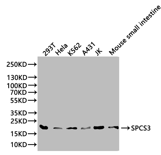
| WB | 1:500-1:2000 |
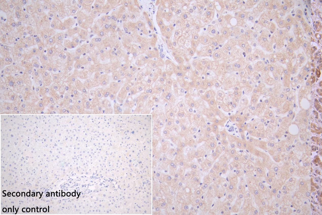
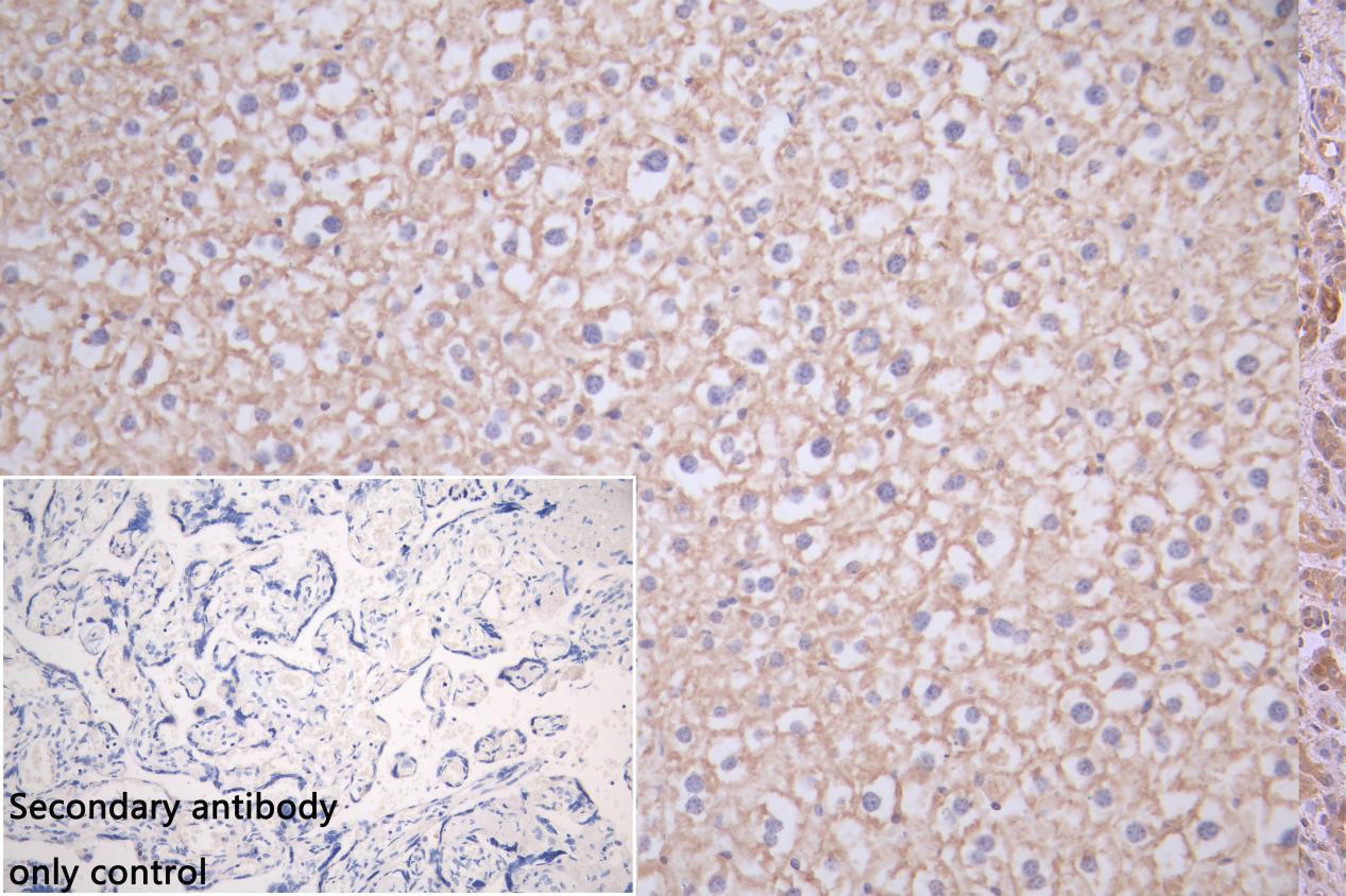
| IHC | 1:20-1:200 |
A rabbit was selected to generate the polyclonal antibody CSB-PA022517LA01HU against SPCS3, using the recombinant human signal peptidase complex subunit 3 protein (33-180aa) as the immunogen. The resulting polyclonal SPCS3 antibody was then purified through antigen affinity purification.
This SPCS3 polyclonal antibody specifically recognizes human and mouse SPCS3 proteins and can be used to detect the endogenous levels of SPCS3 protein. This antibody has undergone quality testing for ELISA, WB, and IHC applications, allowing users to measure, identify, and localize the SPCS3 protein.
There are currently no reviews for this product.

