Mouse Protein disulfide-isomerase(P4HB) ELISA kit

Code CSB-EL017342MO
Size 96T,5×96T,10×96T
Price Request a Quote or Start an on-line Chat
Trial Size 24T ELISA Kit Trial Size (Only USD$150/ kit)
* The sample kit cost can be deducted from your subsequent orders of 96T full size kits of the same analyte at 1/5 per kit, until depleted in 6 months. Apply now

Product Details

Target Name
prolyl 4-hydroxylase, beta polypeptide
Alternative Names
P4hb ELISA Kit; Pdia1Protein disulfide-isomerase ELISA Kit; PDI ELISA Kit; EC 5.3.4.1 ELISA Kit; Cellular thyroid hormone-binding protein ELISA Kit; Endoplasmic reticulum resident protein 59 ELISA Kit; ER protein 59 ELISA Kit; ERp59 ELISA Kit; Prolyl 4-hydroxylase subunit beta ELISA Kit; p55 ELISA Kit
Abbreviation
P4HB
Uniprot No.
Species
Mus musculus (Mouse)
Sample Types
serum, plasma, tissue homogenates
Detection Range
0.312 ng/mL-20 ng/mL
Sensitivity
0.078 ng/mL
Assay Time
1-5h
Sample Volume
50-100ul
Detection Wavelength
450 nm
Research Area
Cancer
Assay Principle
quantitative
Measurement
Sandwich
Precision
Intra-assay Precision (Precision within an assay): CV%<8%
Three samples of known concentration were tested twenty times on one plate to assess.
Inter-assay Precision (Precision between assays): CV%<10%
Three samples of known concentration were tested in twenty assays to assess.
Linearity
To assess the linearity of the assay, samples were spiked with high concentrations of mouse P4HB in various matrices and diluted with the Sample Diluent to produce samples with values within the dynamic range of the assay.
  Sample Serum(n=4)
1:1 Average % 94
Range % 87-98
1:2 Average % 103
Range % 96-108
1:4 Average % 92
Range % 85-97
1:8 Average % 101
Range % 95-107
Recovery
The recovery of mouse P4HB spiked to levels throughout the range of the assay in various matrices was evaluated. Samples were diluted prior to assay as directed in the Sample Preparation section.
Sample Type Average % Recovery Range
Serum (n=5) 89 84-95
EDTA plasma (n=4) 99 91-105
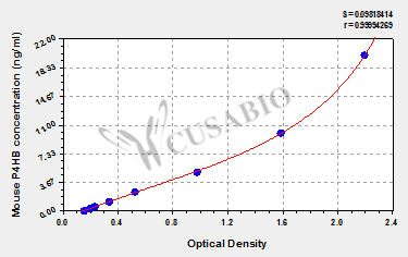
Typical Data
These standard curves are provided for demonstration only. A standard curve should be generated for each set of samples assayed.
ng/ml OD1 OD2 Average Corrected
20 2.248 2.175 2.212 2.044
10 1.615 1.584 1.600 1.432
5 1.016 0.967 0.992 0.824
2.5 0.545 0.536 0.541 0.373
1.25 0.342 0.358 0.350 0.182
0.625 0.251 0.243 0.247 0.079
0.312 0.215 0.210 0.213 0.045
0 0.171 0.165 0.168  
Troubleshooting
and FAQs
Storage
Store at 2-8°C. Please refer to protocol.
Lead Time
3-5 working days after you place the order, and it takes another 3-5 days for delivery via DHL or FedEx
Description

This Mouse P4HB ELISA Kit was designed for the quantitative measurement of Mouse P4HB protein in serum, plasma, tissue homogenates. It is a Sandwich ELISA kit, its detection range is 0.312 ng/mL-20 ng/mL and the sensitivity is 0.078 ng/mL.

Customer Reviews and Q&A

 Customer Reviews

There are currently no reviews for this product.

Submit a Review here

Target Background

Function
(From Uniprot)
This multifunctional protein catalyzes the formation, breakage and rearrangement of disulfide bonds. At the cell surface, seems to act as a reductase that cleaves disulfide bonds of proteins attached to the cell. May therefore cause structural modifications of exofacial proteins. Inside the cell, seems to form/rearrange disulfide bonds of nascent proteins. At high concentrations, functions as a chaperone that inhibits aggregation of misfolded proteins. At low concentrations, facilitates aggregation (anti-chaperone activity). May be involved with other chaperones in the structural modification of the TG precursor in hormone biogenesis. Also acts a structural subunit of various enzymes such as prolyl 4-hydroxylase and microsomal triacylglycerol transfer protein MTTP. Receptor for LGALS9; the interaction retains P4HB at the cell surface of Th2 T helper cells, increasing disulfide reductase activity at the plasma membrane, altering the plasma membrane redox state and enhancing cell migration.
Gene References into Functions
  1. Unfolded protein response caused by protein misfolding, may lead to PDI-dependent NOX activation and contribute to neurotoxicity in neurodegenerative diseases including ALS. PMID: 23118353
  2. PDI from endothelial cells is required for fibrin generation in vivo. PMID: 20668226
  3. Protein disulfide isomerase (PDI) on the platelet surface is recognized by anti-dengue virus NS1 antibodies. PMID: 19822367
Subcellular Location
Endoplasmic reticulum. Endoplasmic reticulum lumen. Melanosome. Cell membrane; Peripheral membrane protein.
Protein Families
Protein disulfide isomerase family
Database Links
icon of phone
Call us
301-363-4651 (Available 9 a.m. to 5 p.m. CST from Monday to Friday)
icon of address
Address
7505 Fannin St., Ste 610, Room 7 (CUBIO Innovation Center), Houston, TX 77054, USA
icon of social media
Join us with

Subscribe newsletter

Leave a message

* To protect against spam, please pass the CAPTCHA test below.
CAPTCHA verification
© 2007-2024 CUSABIO TECHNOLOGY LLC All rights reserved. 鄂ICP备15011166号-1