Recombinant Mouse Cadherin-6 (Cdh6), partial (Active)

In Stock
Code CSB-MP005055MO1
Abbreviation Recombinant Mouse CDH6 protein, partial (Active)
MSDS
Size $116
Order now
Image
  • (Tris-Glycine gel) Discontinuous SDS-PAGE (reduced) with 5% enrichment gel and 15% separation gel.
  • Activity
    Measured by its binding ability in a functional ELISA. Immobilized Mouse CDH6 at 2 μg/ml can bind Anti-CDH6 recombinant antibody (CSB-RA005055MA1HU). The EC50 is 1.884-2.164 ng/mL. Biological Activity Assay
Have Questions? Leave a Message or Start an on-line Chat
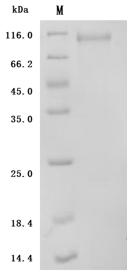
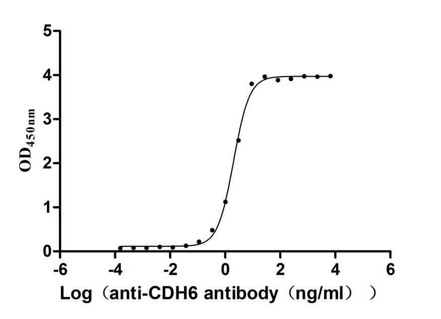
Product Details

Purity
Greater than 95% as determined by SDS-PAGE.
Endotoxin
Less than 1.0 EU/ug as determined by LAL method.
Activity
Measured by its binding ability in a functional ELISA. Immobilized Mouse CDH6 at 2 μg/mL can bind Anti-CDH6 recombinant antibody (CSB-RA005055MA1HU). The EC50 is 1.884-2.164 ng/mL.
Target Names
Uniprot No.
Species
Mus musculus (Mouse)
Source
Mammalian cell
Expression Region
19-615aa
Target Protein Sequence
TFSNPLSKRTSGFPAKRKALELSANSRNELSRSKRSWMWNQFFLLEEYTGSDYQYVGKLHSDQDRGDGSLKYILSGDGAGDLFIINENTGDIQATKRLDREEKPVYILRAQAVNRRTGRPVEPESEFIIKIHDINDNEPIFTKDVYTATVPEMADVGTFVVQVTATDADDPTYGNSAKVVYSILQGQPYFSVESETGIIKTALLNMDRENREQYQVVIQAKDMGGQMGGLSGTTTVNITLTDVNDNPPRFPQSTYQFKTPESSPPGTPIGRIKASDADVGENAEIEYSITDGEGHEMFDVITDQETQEGIITVKKLLDFEKKKVYTLKVEASNPHVEPRFLYLGPFKDSATVRIVVDDVDEPPVFSKLAYILQIREDARINTTIGSVAAQDPDAARNPVKYSVDRHTDMDRIFNIDSGNGSIFTSKLLDRETLLWHNITVIATEINNPKQSSRVPLYIKVLDVNDNAPEFAEFYETFVCEKAKADQLIQTLRAVDKDDPYSGHQFSFSLAPEAASSSNFTIQDNKDNTAGILTRKNGYNRHEMSTYLLPVVISDNDYPVQSSTGTVTVRVCACDHHGNMQSCHAEALIHPTGLSTGA
Mol. Weight
67.8 kDa
Protein Length
Partial
Tag Info
C-terminal 10xHis-tagged
Form
Lyophilized powder
Note: We will preferentially ship the format that we have in stock, however, if you have any special requirement for the format, please remark your requirement when placing the order, we will prepare according to your demand.
Buffer
Lyophilized from a 0.2 μm filtered PBS, 6% Trehalose, pH 7.4
Reconstitution
We recommend that this vial be briefly centrifuged prior to opening to bring the contents to the bottom. Please reconstitute protein in deionized sterile water to a concentration of 0.1-1.0 mg/mL.We recommend to add 5-50% of glycerol (final concentration) and aliquot for long-term storage at -20℃/-80℃. Our default final concentration of glycerol is 50%. Customers could use it as reference.
Troubleshooting and FAQs
Storage Condition
Store at -20°C/-80°C upon receipt, aliquoting is necessary for mutiple use. Avoid repeated freeze-thaw cycles.
Shelf Life
The shelf life is related to many factors, storage state, buffer ingredients, storage temperature and the stability of the protein itself.
Generally, the shelf life of liquid form is 6 months at -20°C/-80°C. The shelf life of lyophilized form is 12 months at -20°C/-80°C.
Lead Time
3-7 business days
Notes
Repeated freezing and thawing is not recommended. Store working aliquots at 4℃ for up to one week.
Datasheet & COA
Please contact us to get it.
Description

This recombinant mouse Cdh6 protein is an active form expressed in a mammalian system to ensure proper folding and native-like functionality. It consists of amino acids 54 to 615 from the mouse Cdh6 sequence and includes a C-terminal 10xHis tag for convenient purification and detection. The recombinant Cdh6 protein exhibits high purity—over 95% as confirmed by SDS-PAGE analysis. Endotoxin content is controlled to be less than 1.0 EU/μg by LAL testing standards. The Cdh6 protein's activity has been validated in a functional ELISA. When coated at 2 μg/mL, it binds specifically to the anti-CDH6 recombinant antibody (CSB-RA005055MA1HU), with an EC50 between 1.884 and 2.164 ng/mL. These features confirm its suitability for applications involving antibody interaction or cadherin-related research.

The mouse Cdh6 protein, a type II cadherin, plays a significant role in various biological processes, particularly in neural development and kidney formation. Its primary functions encompass mediating cell-cell adhesion, contributing to neuronal circuitry, and influencing nephrogenesis.

In the context of the nervous system, Cdh6 is crucial for axon-target matching in retinal ganglion cells (RGCs). Specifically, Cdh6 is expressed by RGCs and their target cells in the visual pathway, facilitating the correct targeting of these axons during development. Studies have demonstrated that Cdh6 acts as a guiding signal that ensures RGCs project appropriately to their synaptic targets [1][2]. Deletion of Cdh6 leads to aberrant projections by RGCs, causing them to target incorrect areas, which could significantly disrupt visual processing [2][3].

Cdh6's involvement is not restricted to neural functions; it also plays a critical role in kidney development. Research has indicated that Cdh6 is vital for the proper formation of nephron structures during embryonic development. Mice lacking this protein exhibit a reduced number of nephrons, attributable to improper epithelial polarization and nephron fusion to ureteric buds [4][5]. This function highlights its essential role in transforming mesenchymal precursors into epithelial cells crucial for nephrogenesis [6].

Additionally, Cdh6 is implicated in various aspects of cellular signaling, particularly during epithelial-mesenchymal transition (EMT), a process critical in both development and cancer metastasis. Cdh6 contributes to regulating intracellular signaling pathways essential for cell adhesion and motility [7][8]. Notably, its expression has been connected to advanced stages of EMT in various cancer types, signifying a dual role in both normal developmental processes and pathological states [9][8].

The interplay of Cdh6 with other cadherins and their associated proteins suggests a complex network of interactions essential for maintaining tissue architecture and function [10]. Cdh6's ability to interact with β-catenin and other cytoplasmic signaling proteins underscores its multifaceted role as both a structural and signaling molecule [11].

References:
[1] J. Osterhout, N. Josten, et al. Cadherin-6 mediates axon-target matching in a non-image-forming visual circuit. Neuron, vol. 71, no. 4, p. 632-639, 2011. https://doi.org/10.1016/j.neuron.2011.07.006
[2] J. Osterhout, R. El‐Danaf, P. Nguyen, & A. Huberman. Birthdate and outgrowth timing predict cellular mechanisms of axon target matching in the developing visual pathway. Cell Reports, vol. 8, no. 4, p. 1006-1017, 2014. https://doi.org/10.1016/j.celrep.2014.06.063
[3] J. Kay, I. Huerta, et al. Retinal ganglion cells with distinct directional preferences differ in molecular identity, structure, and central projections. Journal of Neuroscience, vol. 31, no. 21, p. 7753-7762, 2011. https://doi.org/10.1523/jneurosci.0907-11.2011
[4] N. Treacy, S. Clerkin, et al. Growth and differentiation of human induced pluripotent stem cell (hipsc)-derived kidney organoids using fully synthetic peptide hydrogels. Bioactive Materials, vol. 21, p. 142-156, 2023. https://doi.org/10.1016/j.bioactmat.2022.08.003
[5] U. Dahl, A. Sjödin, et al. Genetic dissection of cadherin function during nephrogenesis. Molecular and Cellular Biology, vol. 22, no. 5, p. 1474-1487, 2002. https://doi.org/10.1128/mcb.22.5.1474-1487.2002
[6] V. Sancisi, G. Gandolfi, et al. Cadherin 6 is a new runx2 target in tgf-β signalling pathway. Plos One, vol. 8, no. 9, p. e75489, 2013. https://doi.org/10.1371/journal.pone.0075489
[7] I. Huerta, I. Kim, P. Voinescu, & J. Sanes. Direction-selective retinal ganglion cells arise from molecularly specified multipotential progenitors. Proceedings of the National Academy of Sciences, vol. 109, no. 43, p. 17663-17668, 2012. https://doi.org/10.1073/pnas.1215806109
[8] M. Gugnoni, V. Sancisi, et al. Cadherin-6 promotes emt and cancer metastasis by restraining autophagy. Oncogene, vol. 36, no. 5, p. 667-677, 2016. https://doi.org/10.1038/onc.2016.237
[9] C. Ma, J. Zhao, et al. Combined overexpression of cadherin 6, cadherin 11 and cluster of differentiation 44 is associated with lymph node metastasis and poor prognosis in oral squamous cell carcinoma. Oncology Letters, 2018. https://doi.org/10.3892/ol.2018.8509
[10] E. Epifanova, В. Салина, et al. Adhesion dynamics in the neocortex determine the start of migration and the post-migratory orientation of neurons. Science Advances, vol. 7, no. 27, 2021. https://doi.org/10.1126/sciadv.abf1973
[11] Q. Liu, M. Dalman, et al. Cell adhesion molecule cadherin‐6 function in zebrafish cranial and lateral line ganglia development. Developmental Dynamics, vol. 240, no. 7, p. 1716-1726, 2011. https://doi.org/10.1002/dvdy.22665

Customer Reviews and Q&A

 Customer Reviews

There are currently no reviews for this product.

Submit a Review here

Target Background

Function
Cadherins are calcium-dependent cell adhesion proteins. They preferentially interact with themselves in a homophilic manner in connecting cells; cadherins may thus contribute to the sorting of heterogeneous cell types.
Gene References into Functions
  1. Cadherin 6 expression in the nascent cortical plate may serve solidification of the protomap for cortical functional areas. PMID: 22875867
  2. cadherin-6 plays essential roles in locomotor activity and ultrasonic vocalization PMID: 23173049
  3. role of cadherin 6 as a novel ligand for alpha(IIb)beta(3) and highlights its function in thrombus formation. PMID: 22539596
  4. Data show that during development, the adhesion molecule cadherin-6 is expressed by a subset of retinal ganglion cells and also by their targets in the brain. PMID: 21867880
  5. Cadherin 6 is expressed in the floor and roof plates of the motor columns of the spinal cord. PMID: 12139922
  6. identified a short 79-bp region that is implicated in regulating expression in cells once they have emigrated from the neural tube PMID: 18234175
  7. Results describe the genetic control of Cdh6 expression by systematic integration of a reporter cassette into bacterial artificial chromosomes, with reporter activities evaluated in stable transgenic mouse lines. PMID: 18617008
  8. These results suggest the role of Cdh6 in coordinating mouse hindbrain compartments possibly under the control of Hox gene regulatory network. PMID: 18948151

Show More

Hide All

Subcellular Location
Cell membrane; Single-pass type I membrane protein.
Database Links
icon of phone
Call us
301-363-4651 (Available 9 a.m. to 5 p.m. CST from Monday to Friday)
icon of address
Address
7505 Fannin St., Ste 610, Room 7 (CUBIO Innovation Center), Houston, TX 77054, USA
icon of social media
Join us with

Subscribe newsletter

Leave a message

* To protect against spam, please pass the CAPTCHA test below.
CAPTCHA verification
© 2007-2025 CUSABIO TECHNOLOGY LLC All rights reserved. 鄂ICP备15011166号-1
Place an order now

I. Product details

*
*
*
*

II. Contact details

*
*

III. Ship To

*
*
*
*
*
*
*

IV. Bill To

*
*
*
*
*
*
*
*