Recombinant Mouse Prolyl endopeptidase FAP (Fap), partial (Active)

In Stock
Code CSB-MP008424MO
Abbreviation Recombinant Mouse Fap protein, partial (Active)
MSDS
Size $190
Order now
Image
  • (Tris-Glycine gel) Discontinuous SDS-PAGE (reduced) with 5% enrichment gel and 15% separation gel.
  • Activity
    Measured by its binding ability in a functional ELISA. Immobilized Mouse Fap at 2 μg/ml can bind Anti-FAP recombinant antibody (CSB-RA008424MA1HU). The EC50 is 0.7918-0.9101 ng/mL. Biological Activity Assay
  • Activity
    FAP Recombinant Monoclonal Antibody (CSB-RA008424MA1HU) captured on Protein A Chip can bind Recombinant Mouse Fap with an affinity constant of 1.51 nM as detected by MetaSPR Assay (WeSPRTM 200). Biological Activity Assay
Have Questions? Leave a Message or Start an on-line Chat
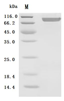
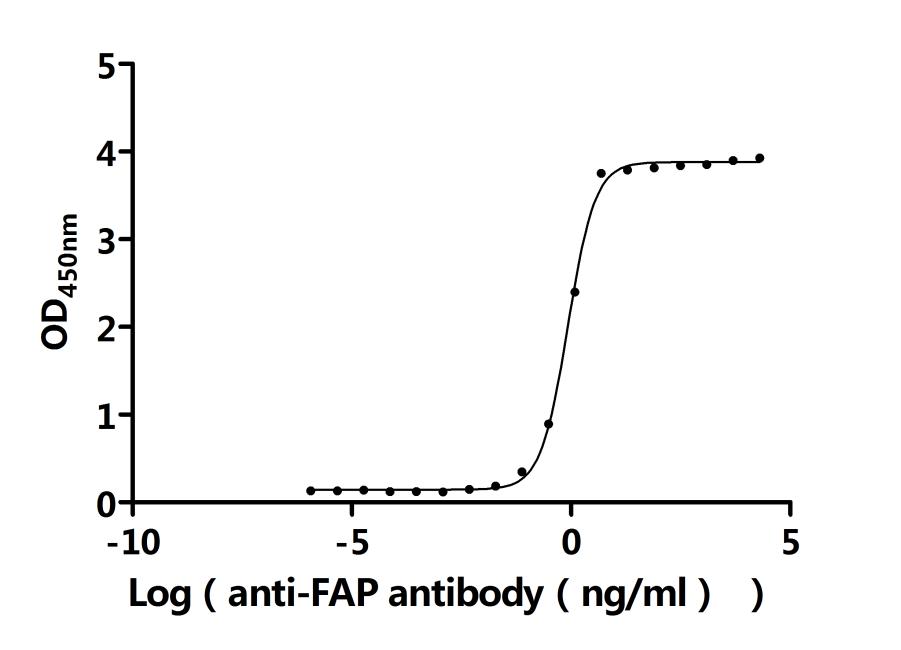
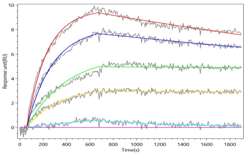
Product Details

Purity
Greater than 95% as determined by SDS-PAGE.
Endotoxin
Less than 1.0 EU/ug as determined by LAL method.
Activity
①Measured by its binding ability in a functional ELISA. Immobilized Mouse Fap at 2 μg/mL can bind Anti-FAP recombinant antibody (CSB-RA008424MA1HU). The EC50 is 0.7918-0.9101 ng/mL.
②FAP Recombinant Monoclonal Antibody (CSB-RA008424MA1HU) captured on Protein A Chip can bind Recombinant Mouse Fap with an affinity constant of 1.51 nM as detected by MetaSPR Assay (WeSPRTM 200).
Target Names
Uniprot No.
Alternative Names
Prolyl endopeptidase FAP; EC 3.4.21.26; Dipeptidyl peptidase FAP; EC 3.4.14.5; Fibroblast activation protein alpha; FAPalpha; Gelatine degradation protease FAP; EC 3.4.21.-; Integral membrane serine protease; Post-proline cleaving enzyme; Serine integral membrane protease; SIMP; Surface-expressed protease; Seprase [Cleaved into: Antiplasmin-cleaving enzyme FAP, soluble form; APCE; EC 3.4.14.5; EC 3.4.21.-; EC 3.4.21.26]
Species
Mus musculus (Mouse)
Source
Mammalian cell
Expression Region
26-761aa
Target Protein Sequence
LRPSRVYKPEGNTKRALTLKDILNGTFSYKTYFPNWISEQEYLHQSEDDNIVFYNIETRESYIILSNSTMKSVNATDYGLSPDRQFVYLESDYSKLWRYSYTATYYIYDLQNGEFVRGYELPRPIQYLCWSPVGSKLAYVYQNNIYLKQRPGDPPFQITYTGRENRIFNGIPDWVYEEEMLATKYALWWSPDGKFLAYVEFNDSDIPIIAYSYYGDGQYPRTINIPYPKAGAKNPVVRVFIVDTTYPHHVGPMEVPVPEMIASSDYYFSWLTWVSSERVCLQWLKRVQNVSVLSICDFREDWHAWECPKNQEHVEESRTGWAGGFFVSTPAFSQDATSYYKIFSDKDGYKHIHYIKDTVENAIQITSGKWEAIYIFRVTQDSLFYSSNEFEGYPGRRNIYRISIGNSPPSKKCVTCHLRKERCQYYTASFSYKAKYYALVCYGPGLPISTLHDGRTDQEIQVLEENKELENSLRNIQLPKVEIKKLKDGGLTFWYKMILPPQFDRSKKYPLLIQVYGGPCSQSVKSVFAVNWITYLASKEGIVIALVDGRGTAFQGDKFLHAVYRKLGVYEVEDQLTAVRKFIEMGFIDEERIAIWGWSYGGYVSSLALASGTGLFKCGIAVAPVSSWEYYASIYSERFMGLPTKDDNLEHYKNSTVMARAEYFRNVDYLLIHGTADDNVHFQNSAQIAKALVNAQVDFQAMWYSDQNHGISSGRSQNHLYTHMTHFLKQCFSLSD
Mol. Weight
91.5 kDa
Protein Length
Partial
Tag Info
C-terminal 10xHis-tagged
Form
Lyophilized powder
Note: We will preferentially ship the format that we have in stock, however, if you have any special requirement for the format, please remark your requirement when placing the order, we will prepare according to your demand.
Buffer
Lyophilized from a 0.2 μm sterile filtered PBS, 6% Trehalose, pH 7.4
Reconstitution
We recommend that this vial be briefly centrifuged prior to opening to bring the contents to the bottom. Please reconstitute protein in deionized sterile water to a concentration of 0.1-1.0 mg/mL.We recommend to add 5-50% of glycerol (final concentration) and aliquot for long-term storage at -20°C/-80°C. Our default final concentration of glycerol is 50%. Customers could use it as reference.
Troubleshooting and FAQs
Storage Condition
Store at -20°C/-80°C upon receipt, aliquoting is necessary for mutiple use. Avoid repeated freeze-thaw cycles.
Shelf Life
The shelf life is related to many factors, storage state, buffer ingredients, storage temperature and the stability of the protein itself.
Generally, the shelf life of liquid form is 6 months at -20°C/-80°C. The shelf life of lyophilized form is 12 months at -20°C/-80°C.
Lead Time
Basically, we can dispatch the products out in 3-7 working days after receiving your orders. Delivery time may differ from different purchasing way or location, please kindly consult your local distributors for specific delivery time.
Notes
Repeated freezing and thawing is not recommended. Store working aliquots at 4℃ for up to one week.
Datasheet & COA
Please contact us to get it.

Customer Reviews and Q&A

 Customer Reviews

There are currently no reviews for this product.

Submit a Review here

Target Background

Function
Cell surface glycoprotein serine protease that participates in extracellular matrix degradation and involved in many cellular processes including tissue remodeling, fibrosis, wound healing, inflammation and tumor growth. Both plasma membrane and soluble forms exhibit post-proline cleaving endopeptidase activity, with a marked preference for Ala/Ser-Gly-Pro-Ser/Asn/Ala consensus sequences, on substrate such as alpha-2-antiplasmin SERPINF2 and SPRY2. Degrade also gelatin, heat-denatured type I collagen, but not native collagen type I and IV, vibronectin, tenascin, laminin, fibronectin, fibrin or casein. Also has dipeptidyl peptidase activity, exhibiting the ability to hydrolyze the prolyl bond two residues from the N-terminus of synthetic dipeptide substrates provided that the penultimate residue is proline, with a preference for Ala-Pro, Ile-Pro, Gly-Pro, Arg-Pro and Pro-Pro. Natural neuropeptide hormones for dipeptidyl peptidase are the neuropeptide Y (NPY), peptide YY (PYY), substance P (TAC1) and brain natriuretic peptide 32 (NPPB). The plasma membrane form, in association with either DPP4, PLAUR or integrins, is involved in the pericellular proteolysis of the extracellular matrix (ECM), and hence promotes cell adhesion, migration and invasion through the ECM. Plays a role in tissue remodeling during development and wound healing. Participates in the cell invasiveness towards the ECM in malignant melanoma cancers. Enhances tumor growth progression by increasing angiogenesis, collagen fiber degradation and apoptosis and by reducing antitumor response of the immune system. Promotes glioma cell invasion through the brain parenchyma by degrading the proteoglycan brevican. Acts as a tumor suppressor in melanocytic cells through regulation of cell proliferation and survival in a serine protease activity-independent manner.
Gene References into Functions
  1. the uptake of the tracer in the joints reflects increased FAP expression during subclinical synovitis and that these joints will show symptoms of inflammation upon disease progression PMID: 29361119
  2. Adora2B stimulation promotes FGF2 and CXCL12 expression in FAP-positive melanoma-associated fibroblasts, contributing to the creation of a tumor-promoting microenvironment. PMID: 27590504
  3. There was no evidence of compensatory upregulation of other DPP4 family members in influenza-infected FAP-deficient mice. FAP appears to be dispensable in anti-influenza adaptive immunity. PMID: 28158223
  4. FAP-STAT3-CCL2 signaling in Cancer-associated fibroblasts (CAF) was sufficient to program an inflammatory component of the tumor microenvironment, which may have particular significance in desmoplasia-associated cancers. PMID: 27216177
  5. this study shows that FAPalpha-targeted modified vaccinia ankara boosting in combination with cyclophosphamide is an effective approach to improving specific anti-tumor immune responses through overcoming immunosuppression PMID: 27545090
  6. Taken together, our study suggested that high FAP expression in CAFs is one reason leading to immune checkpoint blockades resistance in CRC patients and FAP is an optional target for reversing immune checkpoint blockades resistance. PMID: 28302482
  7. FAP-vaccinated mice also treated with Cyclophosphamide chemotherapy showed a marked suppression of tumor growth (inhibition ratio =80%) and a prolongation of survival time. PMID: 28004985
  8. In two different models of pulmonary fibrosis, intratracheal bleomycin instillation and thoracic irradiation, the study finds increased mortality and increased lung fibrosis in FAP-deficient mice compared with wild-type mice. PMID: 26663085
  9. Mouse FGF-21, however, lacks the FAP cleavage site and is not cleaved by FAP. PMID: 26962859
  10. Data indicate that indolamine-2,3-dioxygenase (IDO) and Fibroblast activation protein alpha (FAPalpha) were detectable in B16 melanoma tumor-bearing mice. PMID: 26305550
  11. A transgenic mouse model for pulmonary fibrosis was generated. After bleomycin induction, luciferase cDNA under the control of the FAPa promoter presents strong luminescence in the lungs especially; the expression level reflects the degree of the disease. PMID: 25994578
  12. The FAP(+) stromal cell may have roles in two adverse consequences of cancer. PMID: 23712428
  13. In NIH 3T3 cells overexpressing recombinant mouse FAP, FAP enzymatic activity during matrix production is important for the topographical organization of the ECM fibers. PMID: 21668992
  14. Authors conclude that the proteolytic activity of FAP participates in matrix degradation, but other functions of the protein stimulate increased tumor growth. PMID: 21604185
  15. FAP may be a potentially useful marker for wound age determination. PMID: 20225612
  16. Results suggest that FAP, a product preferentially expressed by TAF, could function as an effective tumor rejection antigen. PMID: 20804499
  17. FAP depletion inhibits tumor cell proliferation indirectly, increases accumulation of collagen, decreases myofibroblast content, and decreases blood vessel density in tumors. PMID: 19920354
  18. the enzymatic activity of fibroblast activation protein plays an important role in the promotion of tumor growth PMID: 15767544
  19. seprase may have a role in promotion of an invasive phenotype by collagenous matrices in ovarian tumor cells PMID: 18823010

Show More

Hide All

Subcellular Location
[Prolyl endopeptidase FAP]: Cell surface. Cell membrane; Single-pass type II membrane protein. Cell projection, lamellipodium membrane; Single-pass type II membrane protein. Cell projection, invadopodium membrane; Single-pass type II membrane protein. Cell projection, ruffle membrane; Single-pass type II membrane protein. Membrane; Single-pass type II membrane protein.; [Antiplasmin-cleaving enzyme FAP, soluble form]: Secreted.
Protein Families
Peptidase S9B family
Tissue Specificity
Expressed strongly in uterus, pancreas, submaxillary gland and skin, less in lymph node, ovary, skeletal muscle, adrenal and bone marrow. Expressed in reactive stromal fibroblast in epithelial cancers. Expressed in melanocytes but not melanomas (at protei
Database Links
CUSABIO guaranteed quality
icon of phone
Call us
301-363-4651 (Available 9 a.m. to 5 p.m. CST from Monday to Friday)
icon of address
Address
7505 Fannin St., Ste 610, Room 7 (CUBIO Innovation Center), Houston, TX 77054, USA
icon of social media
Join us with

Subscribe newsletter

Leave a message

* To protect against spam, please pass the CAPTCHA test below.
CAPTCHA verification
© 2007-2026 CUSABIO TECHNOLOGY LLC All rights reserved. 鄂ICP备15011166号-1
Place an order now

I. Product details

*
*
*
*

II. Contact details

*
*

III. Ship To

*
*
*
*
*
*
*

IV. Bill To

*
*
*
*
*
*
*
*