Full Product Name
Rabbit anti-Homo sapiens (Human) HIST1H3A Polyclonal antibody
Alternative Names
H3 histone family member E pseudogene antibody; H3 histone family; member A antibody; H3/A antibody; H31_HUMAN antibody; H3F3 antibody; H3FA antibody; Hist1h3a antibody; HIST1H3B antibody; HIST1H3C antibody; HIST1H3D antibody; HIST1H3E antibody; HIST1H3F antibody; HIST1H3G antibody; HIST1H3H antibody; HIST1H3I antibody; HIST1H3J antibody; HIST3H3 antibody; histone 1; H3a antibody; Histone cluster 1; H3a antibody; Histone H3 3 pseudogene antibody; Histone H3.1 antibody; Histone H3/a antibody; Histone H3/b antibody; Histone H3/c antibody; Histone H3/d antibody; Histone H3/f antibody; Histone H3/h antibody; Histone H3/i antibody; Histone H3/j antibody; Histone H3/k antibody; Histone H3/l antibody
Species Reactivity
Human, Mouse
Immunogen
Peptide sequence around site of Lys (4) derived from Human Histone H3.1
Immunogen Species
Homo sapiens (Human)
Purification Method
Antigen Affinity Purified
Concentration
It differs from different batches. Please contact us to confirm it.
Buffer
Preservative: 0.03% Proclin 300
Constituents: 50% Glycerol, 0.01M PBS, pH 7.4
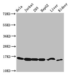
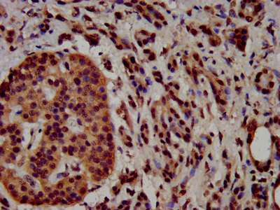
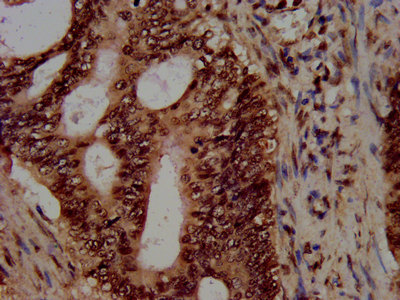
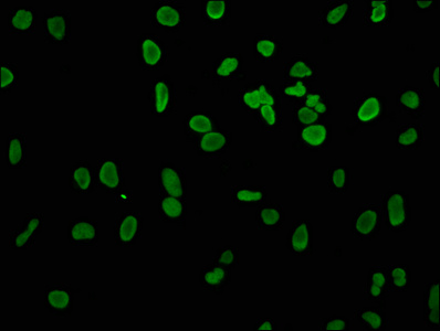
Tested Applications
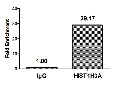
ELISA, WB, IHC, IF, ChIP
Recommended Dilution
| Application |
Recommended Dilution |
| WB |
1:50-1:500 |
| IHC |
1:20-1:200 |
| IF |
1:1-1:10 |
Storage
Upon receipt, store at -20°C or -80°C. Avoid repeated freeze.
Lead Time
Basically, we can dispatch the products out in 1-3 working days after receiving your orders. Delivery time maybe differs from different purchasing way or location, please kindly consult your local distributors for specific delivery time.
Usage
For Research Use Only. Not for use in diagnostic or therapeutic procedures.