MAGOH Antibody

Code CSB-PA013364ESR1HU
Size US$166
Order now
Image
  • Western blot
    All lanes: MAGOH antibody at 4.69μg/ml
    Lane 1: Mouse kidney tissue
    Lane 2: A431 whole cell lysate
    Lane 3: Jurkat whole cell lysate
    Lane 4: Raji whole cell lysate
    Lane 5: K562 whole cell lysate
    Lane 6: Hela whole cell lysate
    Secondary
    Goat polyclonal to rabbit IgG at 1/10000 dilution
    Predicted band size: 18, 13 kDa
    Observed band size: 18 kDa

  • Immunohistochemistry of paraffin-embedded human ovarian cancer using CSB-PA013364ESR1HU at dilution of 1:100

  • Immunohistochemistry of paraffin-embedded human kidney tissue using CSB-PA013364ESR1HU at dilution of 1:100

  • Immunoprecipitating MAGOH in Hela whole cell lysate
    Lane 1: Rabbit control IgG instead of (1μg) instead of CSB-PA013364ESR1HU in Hela whole cell lysate. For western blotting, a HRP-conjugated anti-rabbit IgG, specific to the non-reduced form of IgG was used as the Secondary antibody (1/50000)
    Lane 2: CSB-PA013364ESR1HU (4μg) + Hela whole cell lysate (500μg)
    Lane 3: Hela whole cell lysate (20μg)

Have Questions? Leave a Message or Start an on-line Chat

Product Details

Full Product Name
Rabbit anti-Homo sapiens (Human) MAGOH Polyclonal antibody
Uniprot No.
Target Names
MAGOH
Alternative Names
Mago nashi homolog proliferation associated (Drosophila) antibody; Mago nashi protein homolog antibody; magoh antibody; MAGOHA antibody; MGN_HUMAN antibody; Protein mago nashi homolog antibody
Raised in
Rabbit
Species Reactivity
Human, Mouse
Immunogen
Recombinant Human Protein mago nashi homolog protein (1-146AA)
Immunogen Species
Homo sapiens (Human)
Conjugate
Non-conjugated
Clonality
Polyclonal
Isotype
IgG
Purification Method
Antigen Affinity Purified
Concentration
It differs from different batches. Please contact us to confirm it.
Buffer
PBS with 0.02% sodium azide, 50% glycerol, pH7.3.
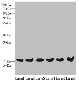
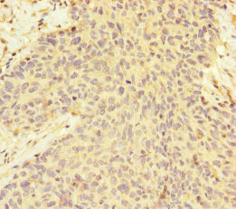
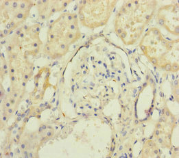
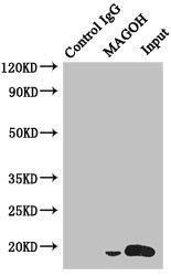
Form
Liquid
Tested Applications
ELISA, WB, IHC, IP
Recommended Dilution
Application Recommended Dilution
WB 1:1000-1:5000
IHC 1:20-1:200
IP 1:200-1:2000
Troubleshooting and FAQs
Storage
Upon receipt, store at -20°C or -80°C. Avoid repeated freeze.
Lead Time
Basically, we can dispatch the products out in 1-3 working days after receiving your orders. Delivery time maybe differs from different purchasing way or location, please kindly consult your local distributors for specific delivery time.

Customer Reviews and Q&A

 Customer Reviews

There are currently no reviews for this product.

Submit a Review here

Target Background

Function
Required for pre-mRNA splicing as component of the spliceosome. Plays a redundant role with MAGOHB as core component of the exon junction complex (EJC) and in the nonsense-mediated decay (NMD) pathway. The EJC is a dynamic structure consisting of core proteins and several peripheral nuclear and cytoplasmic associated factors that join the complex only transiently either during EJC assembly or during subsequent mRNA metabolism. The EJC marks the position of the exon-exon junction in the mature mRNA for the gene expression machinery and the core components remain bound to spliced mRNAs throughout all stages of mRNA metabolism thereby influencing downstream processes including nuclear mRNA export, subcellular mRNA localization, translation efficiency and nonsense-mediated mRNA decay (NMD). The MAGOH-RBM8A heterodimer inhibits the ATPase activity of EIF4A3, thereby trapping the ATP-bound EJC core onto spliced mRNA in a stable conformation. The MAGOH-RBM8A heterodimer interacts with the EJC key regulator PYM1 leading to EJC disassembly in the cytoplasm and translation enhancement of EJC-bearing spliced mRNAs by recruiting them to the ribosomal 48S preinitiation complex. Involved in the splicing modulation of BCL2L1/Bcl-X (and probably other apoptotic genes); specifically inhibits formation of proapoptotic isoforms such as Bcl-X(S); the function is different from the established EJC assembly.
Gene References into Functions
  1. Results provide evidence that MAGOH binding to the C-terminal region of Y14 disrupts Y14 nucleolar localization. PMID: 29330450
  2. findings show 2 genes MAGOH and MAGOHB are expressed in mammals; in macrophages, expression of MAGOHB but not MAGOH mRNA increases after LPS stimulation; both MAGOH proteins interact with other exon junction complex (EJC) components, incorporate into mRNA-bound EJCs and activate nonsense-mediated decay PMID: 23917022
  3. The stable association of multiprotein exon junction complex core with RNA is maintained by inhibition of eIF4AIII ATPase activity by MAGOH-Y14. PMID: 16170325
  4. crystal structure of a tetrameric exon junction core complex containing the DEAD-box adenosine triphosphatase eukaryotic initiation factor 4AIII bound to an ATP analog, MAGOH, Y14, a fragment of MLN51, and a polyuracil mRNA mimic PMID: 16931718
  5. These results indicate that MAGOH regulates the transcriptional activation of STAT3 by interfering complex formation between STAT3 and Y14. PMID: 19254694

Show More

Hide All

Subcellular Location
Nucleus. Nucleus speckle. Cytoplasm.
Protein Families
Mago nashi family
Tissue Specificity
Ubiquitous.
Database Links

HGNC: 6815

OMIM: 602603

KEGG: hsa:4116

STRING: 9606.ENSP00000360525

UniGene: Hs.421576

icon of phone
Call us
301-363-4651 (Available 9 a.m. to 5 p.m. CST from Monday to Friday)
icon of address
Address
7505 Fannin St., Ste 610, Room 7 (CUBIO Innovation Center), Houston, TX 77054, USA
icon of social media
Join us with

Subscribe newsletter

Leave a message

* To protect against spam, please pass the CAPTCHA test below.
CAPTCHA verification
© 2007-2024 CUSABIO TECHNOLOGY LLC All rights reserved. 鄂ICP备15011166号-1
webinars: DT3C facilitates antibody internalization X
Place an order now

I. Product details

*
*
*
*

II. Contact details

*
*

III. Ship To

*
*
*
*
*
*
*

IV. Bill To

*
*
*
*
*
*
*
*