Full Product Name
Rabbit anti-Homo sapiens (Human) MNAT1 Polyclonal antibody
Alternative Names
MNAT 1 antibody; CAP35 antibody; CDK activating kinase assembly factor MAT1 antibody; CDK-activating kinase assembly factor MAT1 antibody; CDK7/cyclin H assembly factor antibody; CDK7/cyclin-H assembly factor antibody; Cyclin G1 interacting protein antibody; Cyclin-G1-interacting protein antibody; MAT1 antibody; MAT1_HUMAN antibody; Menage a trois 1 (CAK assembly factor) antibody; Menage a trois antibody; Menage a trois homolog 1; cyclin H assembly factor (Xenopus laevis) antibody; MNAT CDK activating kinase assembly factor 1 antibody; Mnat1 antibody; p35 antibody; p36 antibody; RING finger protein 66 antibody; RING finger protein MAT1 antibody; RNF66 antibody; TFB3 antibody
Species Reactivity
Human,Mouse,Rat
Immunogen
Synthesized peptide derived from internal of Human MAT1.
Immunogen Species
Homo sapiens (Human)
Purification Method
The antibody was affinity-purified from rabbit antiserum by affinity-chromatography using epitope-specific immunogen.
Concentration
It differs from different batches. Please contact us to confirm it.
Form
Rabbit IgG in phosphate buffered saline (without Mg2+ and Ca2+), pH 7.4, 150mM NaCl, 0.02% sodium azide and 50% glycerol.
Tested Applications
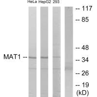
ELISA,WB
Recommended Dilution
| Application |
Recommended Dilution |
| WB |
1:500-1:3000 |
Storage
Upon receipt, store at -20°C or -80°C. Avoid repeated freeze.
Lead Time
Basically, we can dispatch the products out in 1-3 working days after receiving your orders. Delivery time maybe differs from different purchasing way or location, please kindly consult your local distributors for specific delivery time.
Usage
For Research Use Only. Not for use in diagnostic or therapeutic procedures.