OPA1 Antibody

Code CSB-PA016340HA01HU
Size US$166
Order now
Image
  • IHC image of CSB-PA016340HA01HU diluted at 1:300 and staining in paraffin-embedded human small intestine tissue performed on a Leica BondTM system. After dewaxing and hydration, antigen retrieval was mediated by high pressure in a citrate buffer (pH 6.0). Section was blocked with 10% normal goat serum 30min at RT. Then primary antibody (1% BSA) was incubated at 4°C overnight. The primary is detected by a biotinylated secondary antibody and visualized using an HRP conjugated SP system.

  • IHC image of CSB-PA016340HA01HU diluted at 1:300 and staining in paraffin-embedded human heart tissue performed on a Leica BondTM system. After dewaxing and hydration, antigen retrieval was mediated by high pressure in a citrate buffer (pH 6.0). Section was blocked with 10% normal goat serum 30min at RT. Then primary antibody (1% BSA) was incubated at 4°C overnight. The primary is detected by a biotinylated secondary antibody and visualized using an HRP conjugated SP system.

The Latest Promotion Free Antibody trial simple
Have Questions? Leave a Message or Start an on-line Chat

Product Details

Full Product Name
Rabbit anti-Homo sapiens (Human) OPA1 Polyclonal antibody
Uniprot No.
Target Names
OPA1
Alternative Names
OPA1; KIAA0567; Dynamin-like 120 kDa protein, mitochondrial; Optic atrophy protein 1
Raised in
Rabbit
Species Reactivity
Human
Immunogen
Recombinant Human Dynamin-like 120 kDa protein, mitochondrial protein (183-418AA)
Immunogen Species
Homo sapiens (Human)
Conjugate
Non-conjugated

The OPA1 Antibody (Product code: CSB-PA016340HA01HU) is Non-conjugated. For OPA1 Antibody with conjugates, please check the following table.

Available Conjugates
Conjugate Product Code Product Name Application
HRP CSB-PA016340HB01HU OPA1 Antibody, HRP conjugated ELISA
FITC CSB-PA016340HC01HU OPA1 Antibody, FITC conjugated
Biotin CSB-PA016340HD01HU OPA1 Antibody, Biotin conjugated ELISA
Clonality
Polyclonal
Isotype
IgG
Purification Method
>95%, Protein G purified
Concentration
It differs from different batches. Please contact us to confirm it.
Buffer
Preservative: 0.03% Proclin 300
Constituents: 50% Glycerol, 0.01M PBS, pH 7.4
Form
Liquid
Tested Applications
ELISA, IHC
Recommended Dilution
Application Recommended Dilution
IHC 1:200-1:500
Troubleshooting and FAQs
Storage
Upon receipt, store at -20°C or -80°C. Avoid repeated freeze.
Lead Time
Basically, we can dispatch the products out in 1-3 working days after receiving your orders. Delivery time maybe differs from different purchasing way or location, please kindly consult your local distributors for specific delivery time.

Customer Reviews and Q&A

 Customer Reviews

There are currently no reviews for this product.

Submit a Review here

Target Background

Function
Dynamin-related GTPase that is essential for normal mitochondrial morphology by regulating the equilibrium between mitochondrial fusion and mitochondrial fission. Coexpression of isoform 1 with shorter alternative products is required for optimal activity in promoting mitochondrial fusion. Binds lipid membranes enriched in negatively charged phospholipids, such as cardiolipin, and promotes membrane tubulation. The intrinsic GTPase activity is low, and is strongly increased by interaction with lipid membranes. Plays a role in remodeling cristae and the release of cytochrome c during apoptosis. Proteolytic processing in response to intrinsic apoptotic signals may lead to disassembly of OPA1 oligomers and release of the caspase activator cytochrome C (CYCS) into the mitochondrial intermembrane space. Plays a role in mitochondrial genome maintenance.; Inactive form produced by cleavage at S1 position by OMA1 following stress conditions that induce loss of mitochondrial membrane potential, leading to negative regulation of mitochondrial fusion.; Isoforms that contain the alternative exon 4b (present in isoform 4 and isoform 5) are required for mitochondrial genome maintenance, possibly by anchoring the mitochondrial nucleoids to the inner mitochondrial membrane.
Gene References into Functions
  1. Our data indicate that the LEU396ARG mutation in OPA1 is associated with severe dominant optic atrophy. PMID: 29350691
  2. OPA1 gene therapy prevents retinal ganglion cell loss in a dominant optic atrophy mouse model. PMID: 29410463
  3. We have generated a human iPSC line IISHDOi003-A from fibroblasts of a patient with a dominant optic atrophy 'plus' phenotype, harbouring a heterozygous mutation, c.1635C>A; p.Ser545Arg, in the OPA1 gene. PMID: 29034899
  4. OPA1 is a dynamin-related GTPase that controls mitochondrial dynamics, cristae integrity, energetics and mitochondrial DNA maintenance, with eight isoforms being characterized. (Review) PMID: 29382469
  5. Reports an assessment of the afferent visual system and OCT examination in an Italian cohort of fifty-two fully penetrant probands affected by Autosomal Dominant Optic Atrophy (ADOA) with OPA1 mutations and eight asymptomatic carriers of OPA1 mutations. Visual acuity and OCT data in missense mutations were compared with those associated with mutations inducing haploinsufficiency, and correlated with age in both groups. PMID: 29111013
  6. we propose that the SIRT4-OPA1 axis is causally linked to mitochondrial dysfunction and altered mitochondrial dynamics that translates into aging-associated decreased mitophagy based on an unbalanced mitochondrial fusion/fission cycle. PMID: 29081403
  7. Data indicate genotype-phenotype correlations between various types of optic Atrophy 1 (OPA1) mutation and mitophagy. PMID: 28378518
  8. The results show that a metabolic shift from glycolysis in young to mitochondrial respiration in old normal human fibroblasts occurs during chronological lifespan, and MFN1 and OPA1 regulate this process. PMID: 28758339
  9. Genetic testing identified disease-causing mutations in 34% of referred cases, with the majority of these in OPA1. Patients with mutations in OPA1 were more likely to have a family history of disease; however, 30.4% of patients without a family history were also found to have an OPA1 mutation. PMID: 28848318
  10. OPA1 gene mutations were identified in Han Chinese patients with suspected Optic Neuropathy. PMID: 26867657
  11. Identification of genomic rearrangements or pathogenic variants of OPA1 is meaningful for disease prognosis and proper genetic counseling in DOA consultation. PMID: 28668999
  12. In brown adipocytes indirect evidence support the notion that OPA1 regulation of fission serves to increase thermogenesis, which thereby contributes to dissipation of energy. PMID: 28427098
  13. Stabilization of OPA1 impedes cristae remodeling. PMID: 28228254
  14. Our combination of proteomics, biochemistry, genetics, and electron tomography provides a unifying model for mammalian cristae biogenesis by OPA1 and MICOS. PMID: 27974214
  15. The splice site mutation (c.985G>T) identified in the present study led to exon 10 skipping (c.985_1065del, p.V329_D355del), suggesting loss-of-function of the GTPase domain of the OPA1 protein, which is likely to cause haplo-insufficiency, a major disease mechanism in DOA. PMID: 26854526
  16. study identifies a novel pathogenic OPA1 mutation and shows that it is located in the transcript region not prone for NMD activation PMID: 28841713
  17. OPA1 gene screening in patients with bilateral optic atrophy is an important part of clinical evaluation as it may establish correct clinical diagnosis PMID: 27860320
  18. OPA1 and cardiolipin cooperate in heterotypic mitochondrial inner membrane fusion. PMID: 28628083
  19. We propose that OPA1 stabilizes respiratory chain supercomplexes in a conformation that enables respiring mitochondria to compensate a drop in Deltapsim by an explosive matrix pH flash. PMID: 28174208
  20. We report the first cases of genetically confirmed OPA1-related autosomal-dominant optic atrophy from Singapore, including a novel mutation causing 'ADOA plus' syndrome. PMID: 27858935
  21. contrary to conventional notion, S-OPA1 is fully competent for maintaining mitochondrial energetics and cristae structure PMID: 28298442
  22. we analyzed ophthalmological data of a multicentre OPA1 patient cohort and found that women undergo more severe visual loss at adolescence and greater progressive thinning of the retinal nerve fibres than males. Thus, we disclosed a gender-dependent effect on ADOA severity, involving for the first time steroids and Muller glial cells, responsible for RGC degeneration. PMID: 27260406
  23. The architecture of dendritic arborization in patients with OPA1 mutations is not known, but our data support the idea that loss of dendritic arborization may be involved in the pathogenesis of DOA rather than just population loss. PMID: 28125838
  24. OPA1 disclosed a de novo heterozygous deletion c.2012+4_2012+7delAGTA resulting in exon 18 and 19 skipping, which was not detected in healthy family members. PMID: 28245802
  25. This study demonstrated increased mitophagy and excessive mitochondrial fragmentation in primary human cultures associated with DOA plus due to biallelic OPA1 mutations. PMID: 27974645
  26. The present study identified novel compound heterozygous OPA1 mutations in a patient with recessive optic atrophy, sensorimotor neuropathy and congenital cataracts, indicating an expansion of the clinical spectrum of pathologies associated with OPA1 mutations. PMID: 27150940
  27. Optic atrophy type 1, caused by mutations in the OPA1 gene is believed to be the most common hereditary optic neuropathy, and most patients inherit a mutation from an affected parent. In this study we used whole-exome sequencing to investigate the genetic aetiology in a patient affected with isolated optic atrophy. Exome results identified a novel de novo OPA1 mutation. PMID: 27265430
  28. Findings show a new mode of regulation of the mitochondrial fusion proteins, Mfns degradation or OPA1 processing, in response to mitochondrial morphology. PMID: 26935475
  29. Loss of OPA1 protein function by pathogenic OPA1 gene mutation induces increased mitochondrial fragmentation that promotes instability of the mitochondrial respiratory chain complexes. PMID: 27585216
  30. Two heterozygous mutations, p.T414P (c.1240A>C) and p.T540P (c.1618A>C), located in the GTPase and middle domains of OPA1, respectively, were identified in two patients.These two different conformational changes might result in decreased GTPase activities that trigger autosomal dominant optic atrophy associated with auditory neuropathy spectrum disorder PMID: 26905822
  31. A causal link between a pathogenic homozygous OPA1 mutation and hypertrophic cardiomyopathy with optic atrophy was established.It emphasise the vital role played by OPA1 in mitochondrial biogenesis and mtDNA maintenance. PMID: 26561570
  32. OPA1 variants confer risk of leprosy in Chinese Han population and may affect OPA1 expression, mitochondrial function and antimicrobial pathways. PMID: 26360011
  33. Genotype-phenotype heterogeneity in OPA1 autosomal-dominant optic atrophy (ADOA). is evident when inner retinal atrophy is examined as a function of age. PMID: 26385429
  34. Heterozygous mutation in OPA1 disrupts the GTPase domain of OPA1 and is associated with phenotypically variable ADOA Plus. PMID: 26194196
  35. Identification of copy number variation in the gene for autosomal dominant optic atrophy, OPA1, in a Chinese pedigree PMID: 26400325
  36. Data show that OPA1 physiological levels are important in cardiovascular health by maintaining mitochondrial shape and respiratory function, while its down-regulation is associated with cardiovascular disease. [review] PMID: 25557256
  37. increased percentage of apoptotic cells in autosomal dominant optic atrophy patients compared to controls; suggests susceptibility of ADOA cells to oxidative stress and correlation between OPA1 protein dysfunctions and morphological-functional alterations to mitochondria; also results imply sensitivity of mutated protein to free radical damage PMID: 25796301
  38. Distributed abnormalities of diffusivity indexes might reflect abnormal intracellular mitochondrial morphology as well as alteration of protein levels due to OPA1 mutations. PMID: 25794858
  39. A recurrent deletion mutation in OPA1 causes autosomal dominant optic atrophy in a Chinese family. PMID: 25374051
  40. Two heterozygous OPA1 missense mutations affecting highly conserved amino acid positions (p.G488R, p.A495V) were associated with chronic progressive external ophthalmoplegia, parkinsonism, and dementia in two Italian families. PMID: 25820230
  41. OPA1 mutations induced mitochondrial fragmentation, uncoupled mitochondrial respiration, and elicited dysfunctional bioenergetics. PMID: 25744979
  42. The results of this study indicated that underlying the hearing impairment in patients carrying OPA1 missense mutations is a disordered synchrony in auditory nerve fibre activity resulting from neural degeneration affecting the terminal dendrites. PMID: 25564500
  43. Cleavage of the inner membrane fusion factor L-OPA1 is prevented due to the failure to activate the inner membrane protease OMA1 in mitochondria that have a collapsed membrane potential. PMID: 24634514
  44. a novel way in which OPA1 senses energy substrate availability, which modulates its function in the regulation of mitochondrial architecture in a SLC25A protein-dependent manner. PMID: 25298396
  45. OMA1 processing is positively correlated with OPA1 cleavage at the S1 site and the regulation of mitochondrial morphology. PMID: 24719224
  46. The LHON-mtDNA mutations are the most common genetic defects, followed by the OPA1 mutations, in this Chinese cohort. PMID: 25205859
  47. These findings demonstrate that (a) p53 and Oma1 mediate L-Opa1 processing, (b) mitochondrial fragmentation is involved in CDDP-induced apoptosis in OVCA and CECA cells, and (c) dysregulated mitochondrial dynamics PMID: 25112877
  48. we report four cases of children affected by Behr syndrome associated with htetrozygous OPA1 mutation PMID: 25012220
  49. These findings provide additional information regarding the genotype-phenotype correlation and establish the role of the OPA1 gene in Greek patients with autosomal dominant optic atrophy. PMID: 24883014
  50. patients with mutations in the OPA1 gene has shown that about 20% of them present symptoms of a multiple system disease, which may include peripheral neuropathy [review] PMID: 25137924

Show More

Hide All

Involvement in disease
Optic atrophy 1 (OPA1); Dominant optic atrophy plus syndrome (DOA+); Behr syndrome (BEHRS); Mitochondrial DNA depletion syndrome 14, cardioencephalomyopathic type (MTDPS14)
Subcellular Location
Mitochondrion inner membrane; Single-pass membrane protein. Mitochondrion intermembrane space. Mitochondrion membrane.
Protein Families
TRAFAC class dynamin-like GTPase superfamily, Dynamin/Fzo/YdjA family
Tissue Specificity
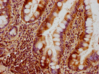
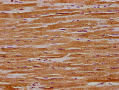
Highly expressed in retina. Also expressed in brain, testis, heart and skeletal muscle. Isoform 1 expressed in retina, skeletal muscle, heart, lung, ovary, colon, thyroid gland, leukocytes and fetal brain. Isoform 2 expressed in colon, liver, kidney, thyr
Database Links

HGNC: 8140

OMIM: 125250

KEGG: hsa:4976

STRING: 9606.ENSP00000354681

UniGene: Hs.594504

icon of phone
Call us
301-363-4651 (Available 9 a.m. to 5 p.m. CST from Monday to Friday)
icon of address
Address
7505 Fannin St., Ste 610, Room 7 (CUBIO Innovation Center), Houston, TX 77054, USA
icon of social media
Join us with

Subscribe newsletter

Leave a message

* To protect against spam, please pass the CAPTCHA test below.
CAPTCHA verification
© 2007-2024 CUSABIO TECHNOLOGY LLC All rights reserved. 鄂ICP备15011166号-1
webinars: DT3C facilitates antibody internalization X
Place an order now

I. Product details

*
*
*
*

II. Contact details

*
*

III. Ship To

*
*
*
*
*
*
*

IV. Bill To

*
*
*
*
*
*
*
*