PACSIN2 Antibody

Code CSB-PA890765EA01HU
Size US$166
Order now
Image
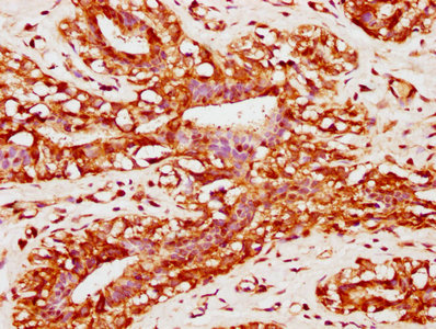
  • IHC image of CSB-PA890765EA01HU diluted at 1:400 and staining in paraffin-embedded human breast cancer performed on a Leica BondTM system. After dewaxing and hydration, antigen retrieval was mediated by high pressure in a citrate buffer (pH 6.0). Section was blocked with 10% normal goat serum 30min at RT. Then primary antibody (1% BSA) was incubated at 4°C overnight. The primary is detected by a biotinylated secondary antibody and visualized using an HRP conjugated SP system.

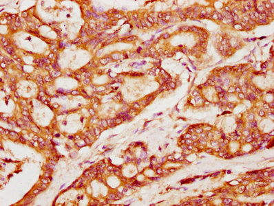
  • IHC image of CSB-PA890765EA01HU diluted at 1:400 and staining in paraffin-embedded human colon cancer performed on a Leica BondTM system. After dewaxing and hydration, antigen retrieval was mediated by high pressure in a citrate buffer (pH 6.0). Section was blocked with 10% normal goat serum 30min at RT. Then primary antibody (1% BSA) was incubated at 4°C overnight. The primary is detected by a biotinylated secondary antibody and visualized using an HRP conjugated SP system.

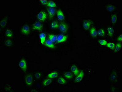
  • Immunofluorescence staining of Hela cells with CSB-PA890765EA01HU at 1:133, counter-stained with DAPI. The cells were fixed in 4% formaldehyde, permeabilized using 0.2% Triton X-100 and blocked in 10% normal Goat Serum. The cells were then incubated with the antibody overnight at 4°C. The secondary antibody was Alexa Fluor 488-congugated AffiniPure Goat Anti-Rabbit IgG(H+L).

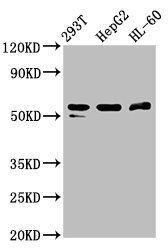
  • Western Blot
    Positive WB detected in: 293T whole cell lysate, HepG2 whole cell lysate, HL60 whole cell lysate
    All lanes: PACSIN2 antibody at 2.6μg/ml
    Secondary
    Goat polyclonal to rabbit IgG at 1/50000 dilution
    Predicted band size: 56, 52 kDa
    Observed band size: 56 kDa

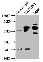
  • Immunoprecipitating PACSIN2 in HepG2 whole cell lysate
    Lane 1: Rabbit control IgG instead of CSB-PA890765EA01HU in HepG2 whole cell lysate. For western blotting, a HRP-conjugated Protein G antibody was used as the secondary antibody (1/2000)
    Lane 2: CSB-PA890765EA01HU (6μg) + HepG2 whole cell lysate (500μg)
    Lane 3: HepG2 whole cell lysate (20μg)

The Latest Promotion Free Antibody trial simple
Have Questions? Leave a Message or Start an on-line Chat

Product Details

Full Product Name
Rabbit anti-Homo sapiens (Human) PACSIN2 Polyclonal antibody
Uniprot No.
Target Names
PACSIN2
Alternative Names
cytoplasmic phosphoprotein PACSIN2 antibody; OTTHUMP00000198281 antibody; OTTHUMP00000198282 antibody; OTTHUMP00000198283 antibody; OTTHUMP00000198284 antibody; PACN2_HUMAN antibody; Pacsin2 antibody; protein kinase C and casein kinase substrate in neurons 2 antibody; Protein kinase C and casein kinase substrate in neurons protein 2 antibody; SDPII antibody; Syndapin II antibody
Raised in
Rabbit
Species Reactivity
Human
Immunogen
Recombinant Human Protein kinase C and casein kinase substrate in neurons protein 2 protein (240-410AA)
Immunogen Species
Homo sapiens (Human)
Conjugate
Non-conjugated

The PACSIN2 Antibody (Product code: CSB-PA890765EA01HU) is Non-conjugated. For PACSIN2 Antibody with conjugates, please check the following table.

Available Conjugates
Conjugate Product Code Product Name Application
HRP CSB-PA890765EB01HU PACSIN2 Antibody, HRP conjugated ELISA
FITC CSB-PA890765EC01HU PACSIN2 Antibody, FITC conjugated
Biotin CSB-PA890765ED01HU PACSIN2 Antibody, Biotin conjugated ELISA
Clonality
Polyclonal
Isotype
IgG
Purification Method
>95%, Protein G purified
Concentration
It differs from different batches. Please contact us to confirm it.
Buffer
Preservative: 0.03% Proclin 300
Constituents: 50% Glycerol, 0.01M PBS, pH 7.4
Form
Liquid
Tested Applications
ELISA, WB, IHC, IF, IP
Recommended Dilution
Application Recommended Dilution
WB 1:500-1:5000
IHC 1:200-1:500
IF 1:50-1:200
IP 1:200-1:2000
Troubleshooting and FAQs
Storage
Upon receipt, store at -20°C or -80°C. Avoid repeated freeze.
Lead Time
Basically, we can dispatch the products out in 1-3 working days after receiving your orders. Delivery time maybe differs from different purchasing way or location, please kindly consult your local distributors for specific delivery time.

Customer Reviews and Q&A

 Customer Reviews

There are currently no reviews for this product.

Submit a Review here

Target Background

Function
Regulates the morphogenesis and endocytosis of caveolae. Lipid-binding protein that is able to promote the tubulation of the phosphatidic acid-containing membranes it preferentially binds. Plays a role in intracellular vesicle-mediated transport. Involved in the endocytosis of cell-surface receptors like the EGF receptor, contributing to its internalization in the absence of EGF stimulus.; (Microbial infection) Specifically enhances the efficiency of HIV-1 virion spread by cell-to-cell transfer. Also promotes the protrusion engulfment during cell-to-cell spread of bacterial pathogens like Listeria monocytogenes. Involved in lipid droplet formation, which is important for HCV virion assembly.
Gene References into Functions
  1. Pacsin2 recruitment is important for maintenance of cell-cell adhesion by stabilizing the VE-cadherin complex within focal adherens junctions and inhibition of its internalization. PMID: 27417273
  2. observations indicate that PACSIN2 promotes the cell-to-cell spreading of HIV-1 by connecting Gag to the actin cytoskeleton. PMID: 29891700
  3. To our knowledge, this is the first study showing PACSIN2 genotype association with hematological toxicity in ALL patients undergoing maintenance therapy. PMID: 27452984
  4. FlnA binding to PACSIN2 F-BAR domain regulates membrane tubulation in megakaryocytes and platelets. PMID: 25838348
  5. our results provide new insights into the mechanism of cellular intoxication by TcdA and the role of PACSIN2 in endocytosis. PMID: 27942025
  6. proposed a packing model for the assembly of PACSIN 2 on membrane, where the proteins are connected by tip-to-tip and wedge loop-mediated lateral interactions on the surface of membrane to generate various diameter tubules PMID: 23888307
  7. PACSIN2 phosphorylation decreases its membrane-binding activity, thereby decreasing its stabilizing effect on caveolae and triggering dynamin-mediated removal of caveolae. PMID: 26092940
  8. PC1 modulates actin cytoskeleton rearrangements and directional cell migration through the Pacsin 2/N-Wasp complex. PMID: 24385601
  9. Cooperation of MICAL-L1, pacsin 2 (syndapin2), and phosphatidic acid in tubular recycling endosome biogenesis. PMID: 23596323
  10. These data indicate that polymorphism in PACSIN2 significantly modulates TPMT activity and influences the risk of GI toxicity associated with mercaptopurine therapy. PMID: 22846425
  11. a novel role for the F-BAR-domain protein PACSIN2 in regulating EGF receptor surface levels and EGF-induced downstream signaling. PMID: 23129763
  12. important role in the formation of plasma membrane caveolae PMID: 21807942
  13. Data identify the BAR-domain protein PACSIN2 as a Rac1 interactor that regulates Rac1-mediated cell spreading and migration. PMID: 21693584
  14. PACSIN2 regulates cell spreading and cell migration, which are dependent on cyclin D1 expression. PMID: 21200149
  15. Results indicate that PACSIN2 mediates membrane sculpting by caveolin-1 in caveola morphology and recruits dynamin-2 for caveola fission. PMID: 21610094
  16. Since the curvature of the neck of the microspike and that of the tubulation share similar geometry, the pacsin2 EFC/F-BAR domain is considered to facilitate both microspike formation and tubulation. PMID: 20188097
  17. Thus, syndapin-dynamin complexes are crucial and sufficient to promote vesicle formation from the trans-Golgi network. PMID: 16551695
  18. These findings suggest a novel function for PACSIN proteins 1, 2, and 3 in dynamic microtubule nucleation. PMID: 18456257
  19. The regulated deformation of membranes and promotion of tubule constrictions by Pacsin suggests a more versatile function of these proteins in vesiculation and endocytosis. PMID: 19549836

Show More

Hide All

Subcellular Location
Cytoplasm. Cytoplasm, cytoskeleton. Cytoplasmic vesicle membrane; Peripheral membrane protein; Cytoplasmic side. Early endosome. Recycling endosome membrane. Cell projection, ruffle membrane; Peripheral membrane protein; Cytoplasmic side. Cell membrane; Peripheral membrane protein; Cytoplasmic side. Cell projection. Membrane, caveola. Note=Detected at the neck of flask-shaped caveolae. Localization to tubular recycling endosomes probably requires interaction with MICALL1 and EHD1.
Protein Families
PACSIN family
Tissue Specificity
Widely expressed.
Database Links

HGNC: 8571

OMIM: 604960

KEGG: hsa:11252

STRING: 9606.ENSP00000263246

UniGene: Hs.162877

icon of phone
Call us
301-363-4651 (Available 9 a.m. to 5 p.m. CST from Monday to Friday)
icon of address
Address
7505 Fannin St., Ste 610, Room 7 (CUBIO Innovation Center), Houston, TX 77054, USA
icon of social media
Join us with

Subscribe newsletter

Leave a message

* To protect against spam, please pass the CAPTCHA test below.
CAPTCHA verification
© 2007-2024 CUSABIO TECHNOLOGY LLC All rights reserved. 鄂ICP备15011166号-1
webinars: DT3C facilitates antibody internalization X
Place an order now

I. Product details

*
*
*
*

II. Contact details

*
*

III. Ship To

*
*
*
*
*
*
*

IV. Bill To

*
*
*
*
*
*
*
*