| Image |
-
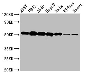
Western Blot
Positive WB detected in: 293T whole cell lysate, U251 whole cell lysate, A549 whole cell lysate, HepG2 whole cell lysate, Hela whole cell lysate, Rat kidney tissue, Mouse heart tissue
All lanes: SLC25A24 antibody at 5µg/ml
Secondary
Goat polyclonal to rabbit IgG at 1/50000 dilution
Predicted band size: 54, 52 kDa
Observed band size: 54 kDa
-
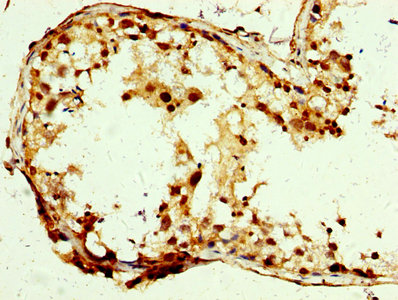
IHC image of CSB-PA743993LA01HU diluted at 1:250 and staining in paraffin-embedded human testis tissue performed on a Leica BondTM system. After dewaxing and hydration, antigen retrieval was mediated by high pressure in a citrate buffer (pH 6.0). Section was blocked with 10% normal goat serum 30min at RT. Then primary antibody (1% BSA) was incubated at 4°C overnight. The primary is detected by a biotinylated secondary antibody and visualized using an HRP conjugated SP system.
-
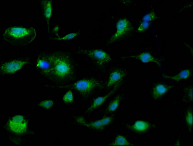
Immunofluorescence staining of U251 cells with CSB-PA743993LA01HU at 1:110, counter-stained with DAPI. The cells were fixed in 4% formaldehyde, permeabilized using 0.2% Triton X-100 and blocked in 10% normal Goat Serum. The cells were then incubated with the antibody overnight at 4°C. The secondary antibody was Alexa Fluor 488-congugated AffiniPure Goat Anti-Rabbit IgG(H+L).
-
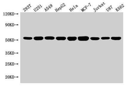
Western Blot
Positive WB detected in: 293T whole cell lysate, U251 whole cell lysate, A549 whole cell lysate, HepG2 whole cell lysate, Hela whole cell lysate, MCF-7 whole cell lysate, Jurkat whole cell lysate, U87 whole cell lysate, K562 whole cell lysate
All lanes: SLC25A24 antibody at 1:1500
Secondary
Goat polyclonal to rabbit IgG at 1/50000 dilution
Predicted band size: 54, 52 kDa
Observed band size: 54 kDa
|