Full Product Name
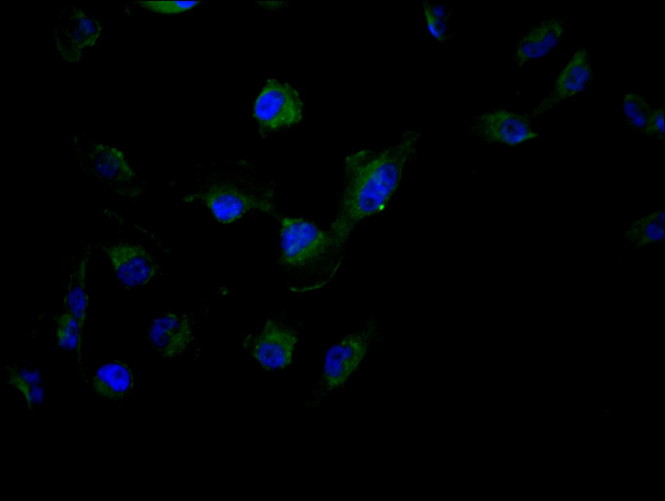
Rabbit anti-Homo sapiens (Human) TRIM4 Polyclonal antibody
Alternative Names
RING finger protein 87 antibody; RNF 87 antibody; RNF87 antibody; TRIM4 antibody; TRIM4_HUMAN antibody; Tripartite motif containing 4 antibody; Tripartite motif protein 4 antibody; Tripartite motif protein TRIM4 antibody; Tripartite motif-containing protein 4 antibody
Immunogen
Recombinant Human E3 ubiquitin-protein ligase TRIM4 protein (1-118AA)
Immunogen Species
Homo sapiens (Human)
Conjugate
Non-conjugated
The TRIM4 Antibody (Product code: CSB-PA866336LA01HU) is Non-conjugated. For TRIM4 Antibody with conjugates, please check the following table.
Available Conjugates
| Conjugate |
Product Code |
Product Name |
Application |
| HRP |
CSB-PA866336LB01HU |
TRIM4 Antibody, HRP conjugated |
ELISA |
| FITC |
CSB-PA866336LC01HU |
TRIM4 Antibody, FITC conjugated |
|
| Biotin |
CSB-PA866336LD01HU |
TRIM4 Antibody, Biotin conjugated |
ELISA |
Purification Method
>95%, Protein G purified
Concentration
It differs from different batches. Please contact us to confirm it.
Buffer
Preservative: 0.03% Proclin 300
Constituents: 50% Glycerol, 0.01M PBS, pH 7.4
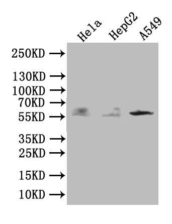
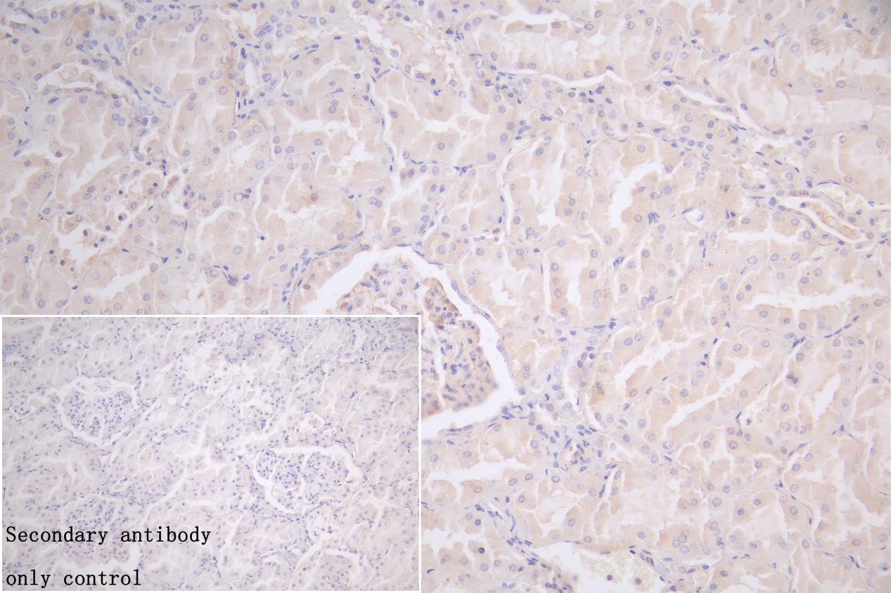
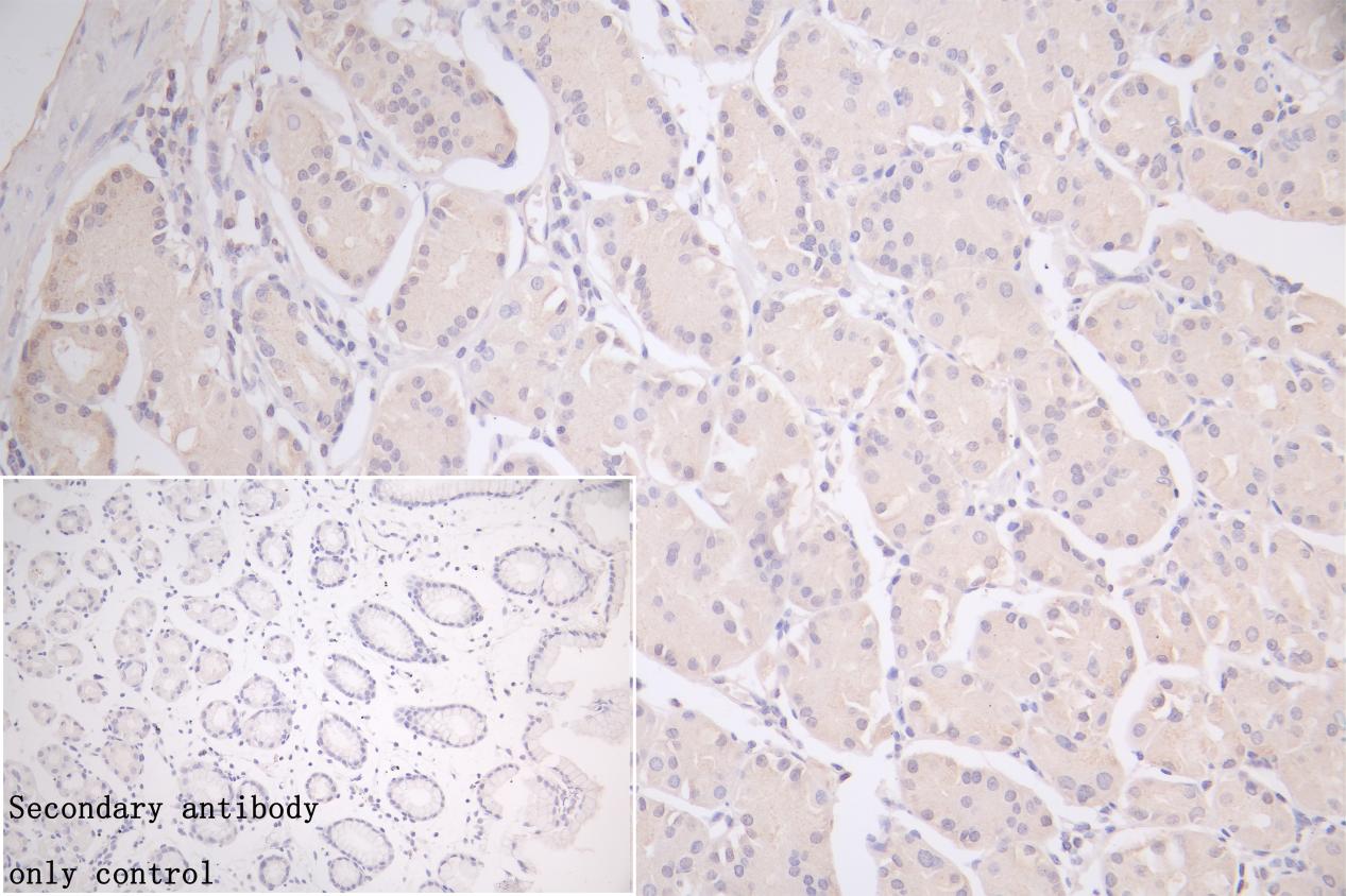
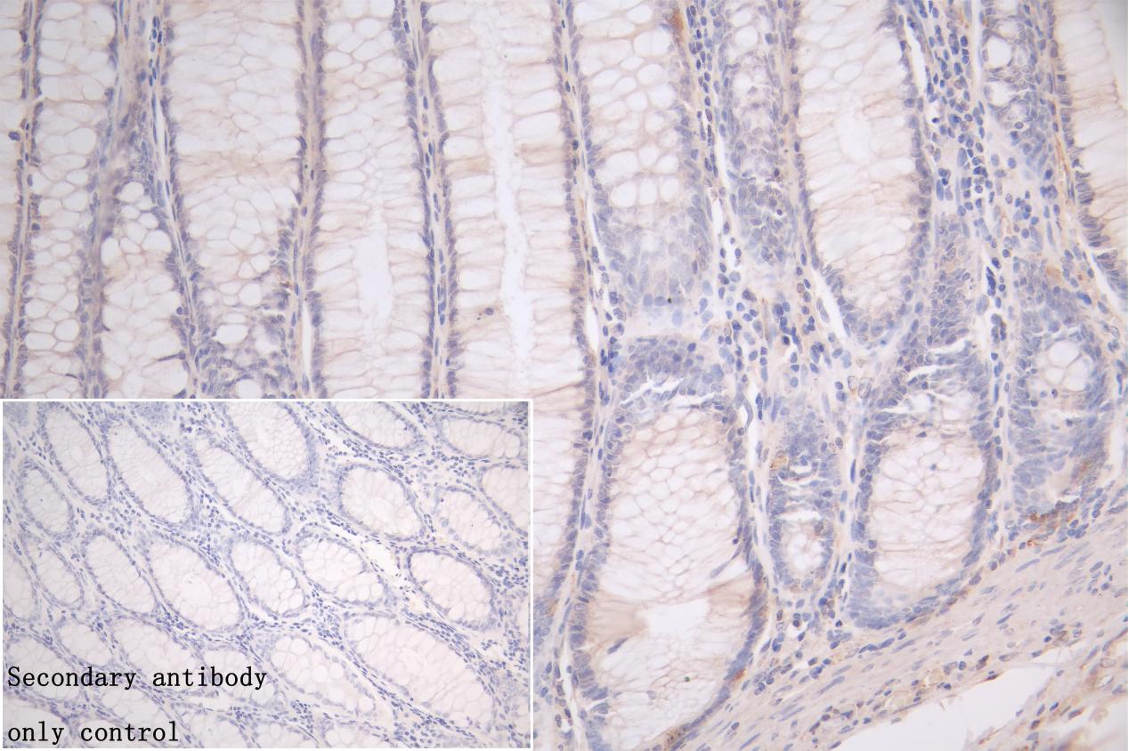
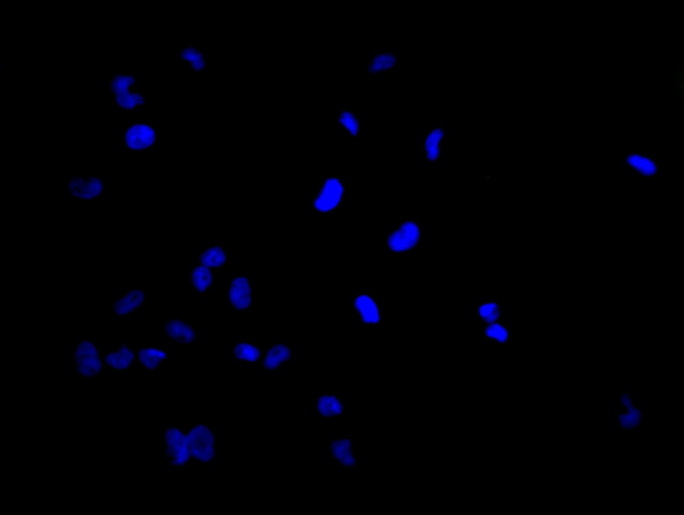
Tested Applications
ELISA, WB, IHC, IF
Recommended Dilution
| Application |
Recommended Dilution |
| WB |
1:500-1:5000 |
| IHC |
1:20-1:200 |
| IF |
1:50-1:200 |
Storage
Upon receipt, store at -20°C or -80°C. Avoid repeated freeze.
Lead Time
Basically, we can dispatch the products out in 1-3 working days after receiving your orders. Delivery time maybe differs from different purchasing way or location, please kindly consult your local distributors for specific delivery time.
Usage
For Research Use Only. Not for use in diagnostic or therapeutic procedures.