Call us
301-363-4651 (Available 9 a.m. to 5 p.m. CST from Monday to Friday)
| Code | CSB-PA836235LA01HU |
| Size | US$166 |
| Order now | |
| Image |
|
| Promotion |  |
| Have Questions? | Leave a Message or Start an on-line Chat |
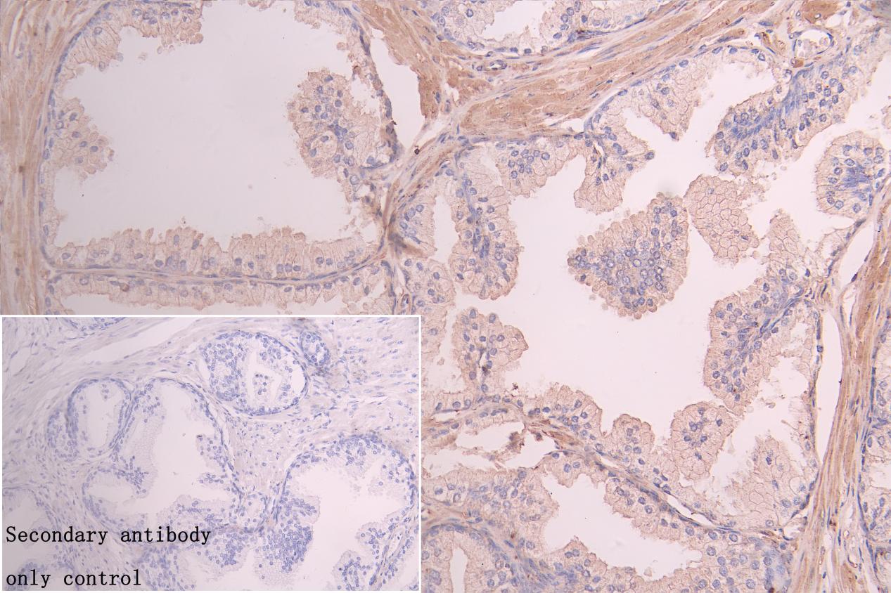
| Application | Recommended Dilution |
|---|---|
| WB | 1:1000-1:3000 |
| IHC | 1:200-1:500 |
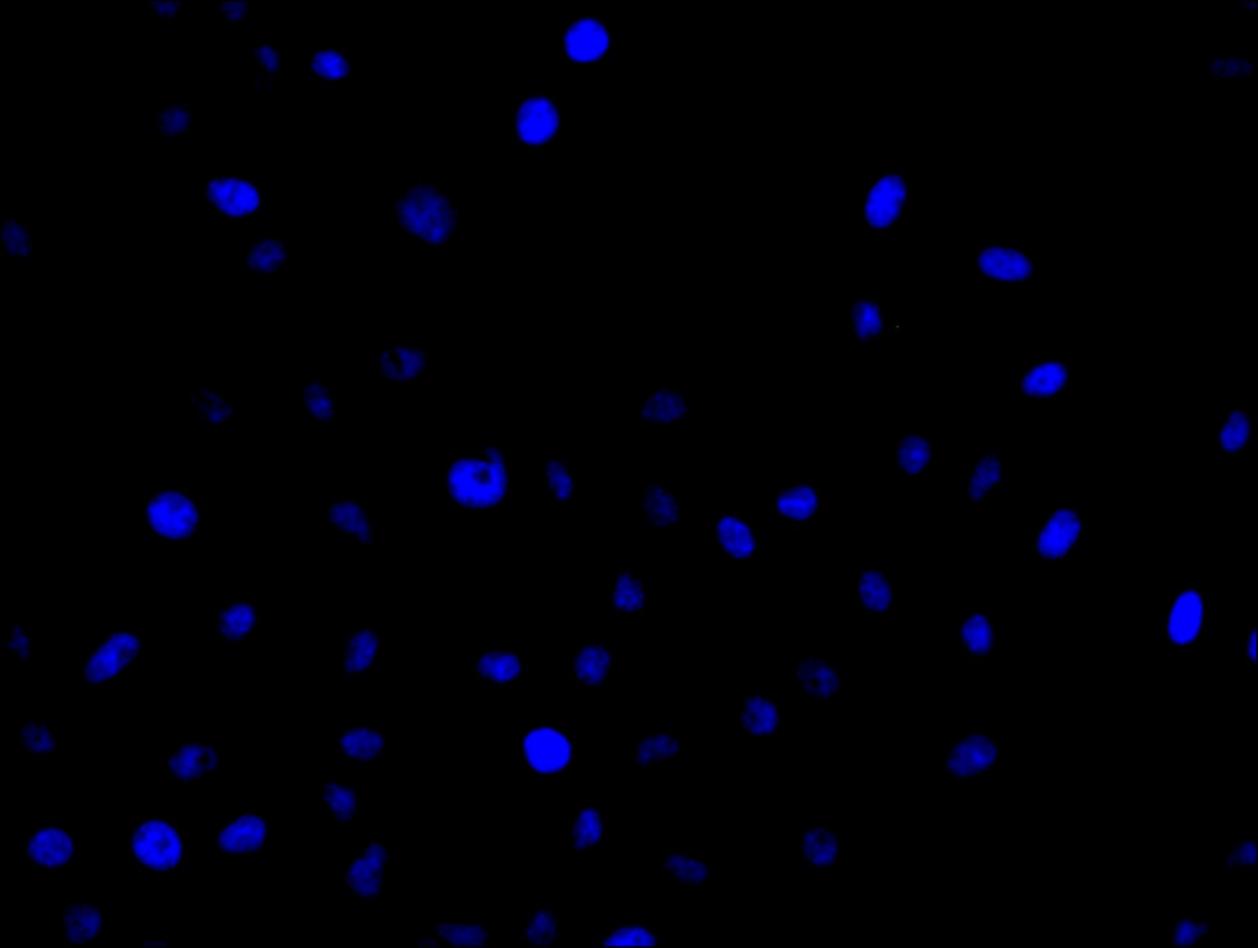
| IF | 1:20-1:100 |
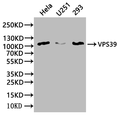
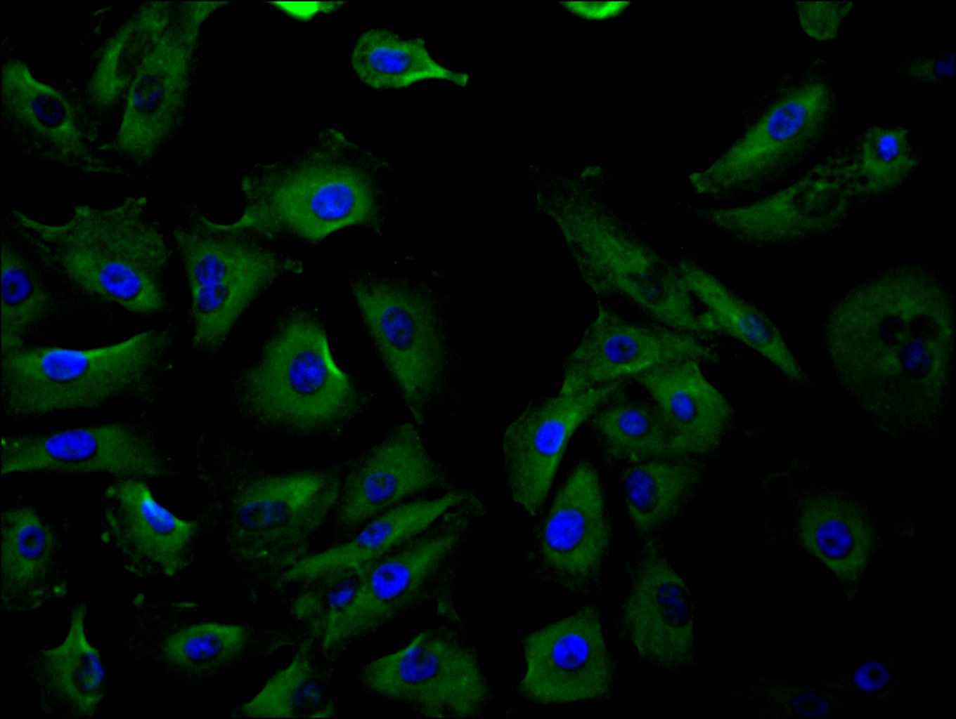
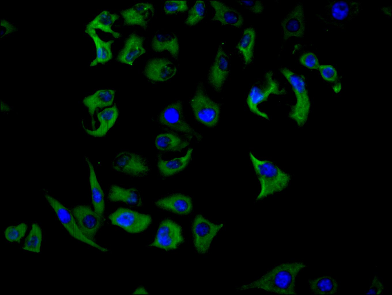
The recombinant peptide corresponding to amino acids 263-490 of the human VPS39 is used as the immunogen to immunize the rabbit to yield the anti-VPS39 antibody. The VPS39 polyclonal antibody exists as an unconjugated IgG isoform. Its purity is 95%+ using protein G purified. It reacts with human and mouse VPS39. This VPS39 antibody has been validated for use in ELISA, WB, IF, and IHC analyses.
VPS39 mainly regulates endosomal membrane trafficking and fusion. It is a subunit of the homotypic fusion and protein sorting (HOPS) complex, which is involved in the tethering and fusion of endosomes and lysosomes. It is also involved in the regulation of lysosomal biogenesis, autophagy, and signaling pathways. Mutations in the VPS39 gene have been linked to various diseases such as Hermansky-Pudlak syndrome.
There are currently no reviews for this product.

