Call us
301-363-4651 (Available 9 a.m. to 5 p.m. CST from Monday to Friday)
| Code | CSB-MP005055HU1 |
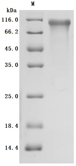
| Abbreviation | Recombinant Human CDH6 protein, partial (Active) |
| MSDS | |
| Size | $104 |
| Order now | |
| Image |
|
| Have Questions? | Leave a Message or Start an on-line Chat |
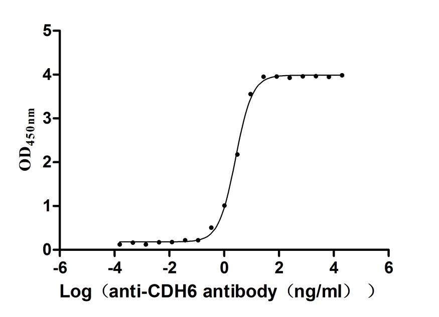
Cadherin-6 mediates calcium-dependent cell–cell adhesion in renal and neural tissues, and its aberrant expression correlates with metastatic potential in multiple cancer types. This recombinant fragment spans residues 19–615, encompassing the extracellular cadherin repeats critical for homophilic binding, and demonstrates specific antibody recognition with an EC50 of 2.421–2.802 ng/mL in functional ELISA, confirming native epitope presentation. The measured binding activity supports use in antibody development and validation workflows, as well as in cell adhesion and migration assays where cadherin-mediated interactions drive experimental readouts. Produced in mammalian cells with greater than 95% purity and endotoxin levels below 1.0 EU/μg, this protein meets the quality thresholds commonly required for tumor metastasis studies and functional assays demanding authentic post-translational modifications.
There are currently no reviews for this product.

