Recombinant Mouse Complement factor I (Cfi)

In Stock Promotion
Code CSB-MP005279MO
MSDS
Size $660
Promotion
A Special Offer on CUSABIO in Stock Proteins
Order now
Image
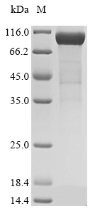
  • (Tris-Glycine gel) Discontinuous SDS-PAGE (reduced) with 5% enrichment gel and 15% separation gel.
Have Questions? Leave a Message or Start an on-line Chat

Product Details

Purity
Greater than 85% as determined by SDS-PAGE.
Target Names
Uniprot No.
Research Area
Epigenetics and Nuclear Signaling
Alternative Names
Cfi; IfComplement factor I; EC 3.4.21.45; C3B/C4B inactivator) [Cleaved into: Complement factor I heavy chain; Complement factor I light chain]
Species
Mus musculus (Mouse)
Source
Mammalian cell
Expression Region
19-603aa
Target Protein Sequence
KNTPASGQPQEDLVEQKCLLKNYTHHSCDKVFCQPWQKCIEGTCACKLPYQCPKAGTPVCATNGRGYPTYCHLKSFECLHPEIKFSNNGTCTAEEKFNVSLIYGSTDTEGIVQVKLVDQDEKMFICKNSWSTVEANVACFDLGFPLGVRDIQGRFNIPVNHKINSTECLHVRCQGVETSLAECTFTKKSSKAPHGLAGVVCYTQDADFPTSQSFQCVNGKRIPQEKACDGVNDCGDQSDELCCKGCRGQAFLCKSGVCIPNQRKCNGEVDCITGEDESGCEEDKKNKIHKGLARSDQGGETEIETEETEMLTPDMDTERKRIKSLLPKLSCGVKRNTHIRRKRVVGGKPAEMGDYPWQVAIKDGDRITCGGIYIGGCWILTAAHCVRPSRYRNYQVWTSLLDWLKPNSQLAVQGVSRVVVHEKYNGATYQNDIALVEMKKHPGKKECELINSVPACVPWSPYLFQPNDRCIISGWGREKDNQKVYSLRWGEVDLIGNCSRFYPGRYYEKEMQCAGTSDGSIDACKGDSGGPLVCKDVNNVTYVWGIVSWGENCGKPEFPGVYTRVASYFDWISYYVGRPLVSQYNV
Note: The complete sequence including tag sequence, target protein sequence and linker sequence could be provided upon request.
Mol. Weight
69.8 kDa
Protein Length
Full Length of Mature Protein
Tag Info
N-terminal 10xHis-tagged and C-terminal Myc-tagged
Form
Liquid or Lyophilized powder
Note: We will preferentially ship the format that we have in stock, however, if you have any special requirement for the format, please remark your requirement when placing the order, we will prepare according to your demand.
Buffer
If the delivery form is liquid, the default storage buffer is Tris/PBS-based buffer, 5%-50% glycerol. If the delivery form is lyophilized powder, the buffer before lyophilization is Tris/PBS-based buffer, 6% Trehalose.
Reconstitution
We recommend that this vial be briefly centrifuged prior to opening to bring the contents to the bottom. Please reconstitute protein in deionized sterile water to a concentration of 0.1-1.0 mg/mL.We recommend to add 5-50% of glycerol (final concentration) and aliquot for long-term storage at -20°C/-80°C. Our default final concentration of glycerol is 50%. Customers could use it as reference.
Troubleshooting and FAQs
Storage Condition
Store at -20°C/-80°C upon receipt, aliquoting is necessary for mutiple use. Avoid repeated freeze-thaw cycles.
Shelf Life
The shelf life is related to many factors, storage state, buffer ingredients, storage temperature and the stability of the protein itself.
Generally, the shelf life of liquid form is 6 months at -20°C/-80°C. The shelf life of lyophilized form is 12 months at -20°C/-80°C.
Lead Time
3-7 business days
Notes
Repeated freezing and thawing is not recommended. Store working aliquots at 4°C for up to one week.
Datasheet & COA
Please contact us to get it.
Description

Complement factor I (CFI) is an 88 kDa glycoprotein primarily synthesized by the liver [1]. It is a serine protease that plays a crucial role in regulating the complement system activation [2]. CFI functions as a complement inhibitor, deactivating C3b and C4 to prevent the formation of C3 convertase, thereby negatively regulating the alternative and classical complement pathways [3]. CFI achieves this by degrading C4b and C3b in the presence of cofactors such as C4b-binding protein (C4BP), complement factor H (CFH), membrane cofactor protein (MCP), and complement receptor 1 (CR1) [4]. Mutations in the CFI gene have been associated with atypical hemolytic uremic syndrome (aHUS), leading to altered secretion or function of CFI [5]. Additionally, CFI has been implicated in age-related macular degeneration (AMD), with rare variants within CFI being reported in advanced AMD patients [6].

Furthermore, genetic abnormalities in CFI, along with other complement regulatory proteins, have been linked to aHUS, with more than 120 mutations in CFH, CFI, and MCP reported in aHUS patients [7]. These genetic variants in the complement system, including CFI, account for a significant proportion of aHUS cases [8]. Moreover, CFI, along with other complement regulators, has been found to have a relative role in both sporadic and familial aHUS, impacting the clinical phenotype of the disease [7].

References:
[1] A. Shields, A. Pagnamenta, P. Aj, J. Taylor, H. Allroggen, S. Patelet al., "Classical and non-classical presentations of complement factor i deficiency: two contrasting cases diagnosed via genetic and genomic methods", Frontiers in Immunology, vol. 10, 2019. https://doi.org/10.3389/fimmu.2019.01150
[2] Y. Liu, Z. Lv, T. Xiao, X. Zhang, C. Ding, B. Qinet al., "Functional and expressional analyses reveal the distinct role of complement factor i in regulating complement system activation during gcrv infection in ctenopharyngodon idella", International Journal of Molecular Sciences, vol. 23, no. 19, p. 11369, 2022. https://doi.org/10.3390/ijms231911369
[3] E. Levit, J. Leon, & M. Lincoln, "Pearls & oy-sters: homozygous complement factor i deficiency presenting as fulminant relapsing complement-mediated cns vasculitis", Neurology, vol. 101, no. 2, 2023. https://doi.org/10.1212/wnl.0000000000207079
[4] X. Cai, W. Qiu, M. Qian, S. Feng, C. Peng, J. Zhanget al., "A candidate prognostic biomarker complement factor i promotes malignant progression in glioma", Frontiers in Cell and Developmental Biology, vol. 8, 2021. https://doi.org/10.3389/fcell.2020.615970
[5] S. Nilsson, N. Kalchishkova, L. Trouw, V. Frémeaux‐Bacchi, B. Villoutreix, & A. Blom, "Mutations in complement factor i as found in atypical hemolytic uremic syndrome lead to either altered secretion or altered function of factor i", European Journal of Immunology, vol. 40, no. 1, p. 172-185, 2009. https://doi.org/10.1002/eji.200939280
[6] D. Qian, M. Kan, X. Weng, H. Yugeng, C. Zhou, Y. Gen-longet al., "Common variant rs10033900 near the complement factor i gene is associated with age-related macular degeneration risk in han chinese population", European Journal of Human Genetics, vol. 22, no. 12, p. 1417-1419, 2014. https://doi.org/10.1038/ejhg.2014.37
[7] M. Noris, J. Caprioli, E. Bresin, C. Mossali, G. Pianetti, S. Gambaet al., "Relative role of genetic complement abnormalities in sporadic and familial ahus and their impact on clinical phenotype", Clinical Journal of the American Society of Nephrology, vol. 5, no. 10, p. 1844-1859, 2010. https://doi.org/10.2215/cjn.02210310
[8] M. Geerlings, E. Volokhina, N. Kar, M. Pauper, C. Hoyng, L. Heuvelet al., "Genotype‐phenotype correlations of low‐frequency variants in the complement system in renal disease and age‐related macular degeneration", Clinical Genetics, vol. 94, no. 3-4, p. 330-338, 2018. https://doi.org/10.1111/cge.13392

Customer Reviews and Q&A

 Customer Reviews

There are currently no reviews for this product.

Submit a Review here

Target Background

Function
Trypsin-like serine protease that plays an essential role in regulating the immune response by controlling all complement pathways. Inhibits these pathways by cleaving three peptide bonds in the alpha-chain of C3b and two bonds in the alpha-chain of C4b thereby inactivating these proteins. Essential cofactors for these reactions include factor H and C4BP in the fluid phase and membrane cofactor protein/CD46 and CR1 on cell surfaces. The presence of these cofactors on healthy cells allows degradation of deposited C3b by CFI in order to prevent undesired complement activation, while in apoptotic cells or microbes, the absence of such cofactors leads to C3b-mediated complement activation and subsequent opsonization.
Gene References into Functions
  1. Factor I-mediated generation of activated C3 fragments in the circulation is a critical determinant for the development of MPGN2 associated with factor H deficiency. PMID: 18202746
Subcellular Location
Secreted, extracellular space.
Protein Families
Peptidase S1 family
Tissue Specificity
Expressed in the liver by hepatocytes. Also present in other cells such as monocytes, fibroblasts or keratinocytes.
Database Links
icon of phone
Call us
301-363-4651 (Available 9 a.m. to 5 p.m. CST from Monday to Friday)
icon of address
Address
7505 Fannin St., Ste 610, Room 7 (CUBIO Innovation Center), Houston, TX 77054, USA
icon of social media
Join us with

Subscribe newsletter

Leave a message

* To protect against spam, please pass the CAPTCHA test below.
CAPTCHA verification
© 2007-2024 CUSABIO TECHNOLOGY LLC All rights reserved. 鄂ICP备15011166号-1
Place an order now

I. Product details

*
*
*
*

II. Contact details

*
*

III. Ship To

*
*
*
*
*
*
*

IV. Bill To

*
*
*
*
*
*
*
*