AP2A1 Recombinant Monoclonal Antibody

Code CSB-RA191307A0HU
Size US$210
Order now
Image
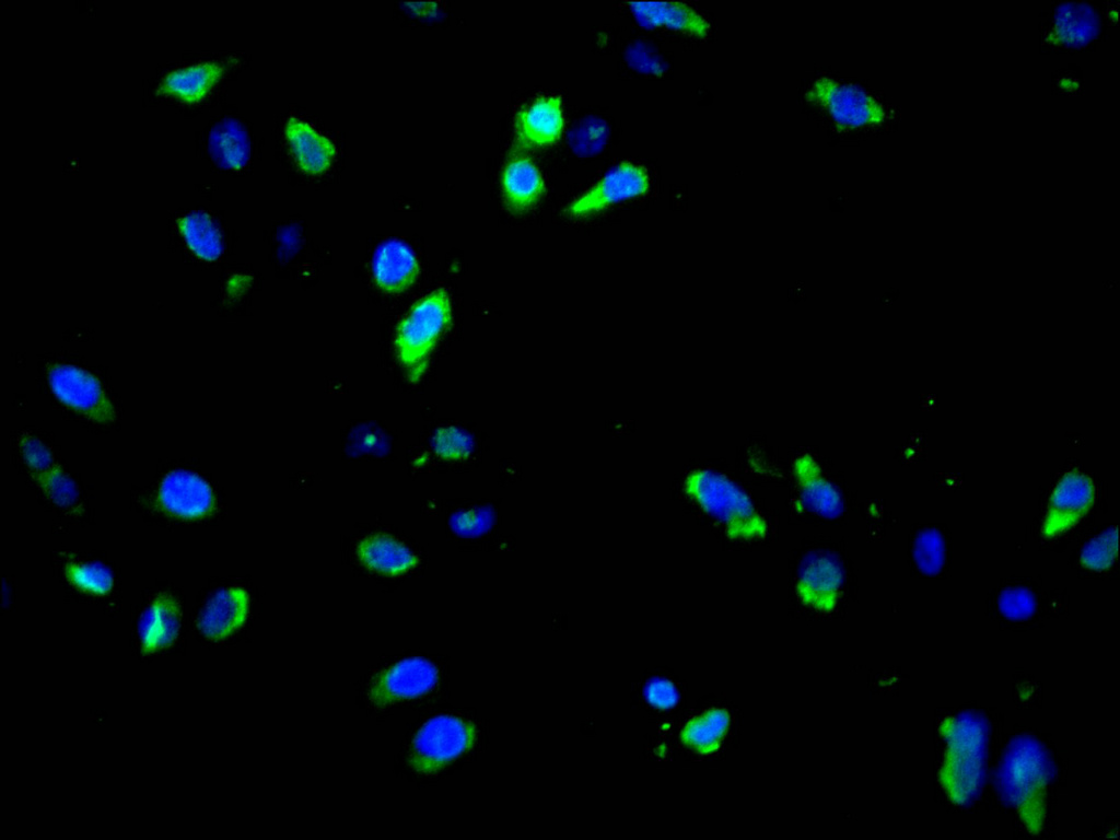
  • Immunofluorescence staining of Hela cell with CSB-RA191307A0HU at 1:50, counter-stained with DAPI. The cells were fixed in 4% formaldehyde and blocked in 10% normal Goat Serum. The cells were then incubated with the antibody overnight at 4°C. The secondary antibody was Alexa Fluor 488-congugated AffiniPure Goat Anti-Rabbit IgG(H+L).
Have Questions? Leave a Message or Start an on-line Chat

Product Details

Uniprot No.
Target Names
Alternative Names
AP-2 complex subunit alpha-1 (100 kDa coated vesicle protein A) (Adaptor protein complex AP-2 subunit alpha-1) (Adaptor-related protein complex 2 subunit alpha-1) (Alpha-adaptin A) (Alpha1-adaptin) (Clathrin assembly protein complex 2 alpha-A large chain) (Plasma membrane adaptor HA2/AP2 adaptin alpha A subunit), AP2A1, ADTAA CLAPA1
Species Reactivity
Human
Immunogen
A synthesized peptide from human AP2A1 protein
Immunogen Species
Homo sapiens (Human)
Conjugate
Non-conjugated
Clonality
Monoclonal
Isotype
Rabbit IgG
Clone No.
4D7
Purification Method
Affinity-chromatography
Concentration
It differs from different batches. Please contact us to confirm it.
Buffer
Rabbit IgG in 10mM phosphate buffered saline , pH 7.4, 150mM sodium chloride, 0.05% BSA, 0.02% sodium azide and 50% glycerol.
Form
Liquid
Tested Applications
ELISA, IF
Recommended Dilution
Application Recommended Dilution
IF 1:50-1:200
Troubleshooting and FAQs
Storage
Upon receipt, store at -20°C or -80°C. Avoid repeated freeze.
Lead Time
Basically, we can dispatch the products out in 1-3 working days after receiving your orders. Delivery time maybe differs from different purchasing way or location, please kindly consult your local distributors for specific delivery time.
Description

AP2A1, also known as alpha1-adaptin, serves as a critical component of the AP-2 adaptor protein complex, which orchestrates clathrin-mediated endocytosis at the plasma membrane. This process is fundamental to receptor internalization, nutrient uptake, and synaptic vesicle recycling, making AP2A1 an essential target for researchers investigating membrane trafficking, signal transduction, and neurobiology.

This recombinant monoclonal antibody, clone 4D7, offers the reproducibility and consistency that demanding experimental workflows require. Generated against a synthetic peptide derived from human AP2A1, the antibody is produced using recombinant technology, ensuring sequence-defined specificity and eliminating the lot-to-lot variability often encountered with traditional hybridoma-derived antibodies. This reliability is particularly valuable for longitudinal studies or when establishing standardized protocols across research teams.

The antibody has been validated for immunofluorescence applications, with testing performed on HeLa cells demonstrating clear cytoplasmic staining patterns consistent with AP2A1's known localization at the plasma membrane and clathrin-coated pits. Working dilutions of 1:50 to 1:200 provide flexibility for optimization across different experimental conditions and imaging systems. The validation protocol employed standard fixation with 4% formaldehyde and blocking with normal goat serum, followed by overnight primary antibody incubation and detection with Alexa Fluor 488-conjugated secondary antibody.

Supplied in a glycerol-containing buffer optimized for long-term stability, this antibody supports research into endocytic pathways, receptor trafficking dynamics, and clathrin-dependent cellular processes. It represents a valuable tool for cell biologists exploring membrane dynamics and investigators studying how disrupted endocytosis contributes to disease states.

Usage
For Research Use Only. Not for use in diagnostic or therapeutic procedures.

Customer Reviews and Q&A

 Customer Reviews

There are currently no reviews for this product.

Submit a Review here

Target Background

Function
Component of the adaptor protein complex 2 (AP-2). Adaptor protein complexes function in protein transport via transport vesicles in different membrane traffic pathways. Adaptor protein complexes are vesicle coat components and appear to be involved in cargo selection and vesicle formation. AP-2 is involved in clathrin-dependent endocytosis in which cargo proteins are incorporated into vesicles surrounded by clathrin (clathrin-coated vesicles, CCVs) which are destined for fusion with the early endosome. The clathrin lattice serves as a mechanical scaffold but is itself unable to bind directly to membrane components. Clathrin-associated adaptor protein (AP) complexes which can bind directly to both the clathrin lattice and to the lipid and protein components of membranes are considered to be the major clathrin adaptors contributing the CCV formation. AP-2 also serves as a cargo receptor to selectively sort the membrane proteins involved in receptor-mediated endocytosis. AP-2 seems to play a role in the recycling of synaptic vesicle membranes from the presynaptic surface. AP-2 recognizes Y-X-X-[FILMV] (Y-X-X-Phi) and [ED]-X-X-X-L-[LI] endocytosis signal motifs within the cytosolic tails of transmembrane cargo molecules. AP-2 may also play a role in maintaining normal post-endocytic trafficking through the ARF6-regulated, non-clathrin pathway. During long-term potentiation in hippocampal neurons, AP-2 is responsible for the endocytosis of ADAM10. The AP-2 alpha subunit binds polyphosphoinositide-containing lipids, positioning AP-2 on the membrane. The AP-2 alpha subunit acts via its C-terminal appendage domain as a scaffolding platform for endocytic accessory proteins. The AP-2 alpha and AP-2 sigma subunits are thought to contribute to the recognition of the [ED]-X-X-X-L-[LI] motif.
Gene References into Functions
  1. Results revealed thirteen novel splice junctions of previously annotated exons. Consecutive nested PCRs led to the determination of the primary structure of seventy-seven novel AP2A1 transcripts, all of which were shown to comprise at least one premature translation termination codon, thus representing nonsense-mediated mRNA decay candidates. PMID: 30081190
  2. AP-2alpha depletion enhanced apoptosis, demonstrating that disrupting the HIV-1 Nef:AP-2alpha interaction limits Nef-mediated apoptosis. PMID: 28577469
  3. These results lead to a model for the docking of the full AP-2 tetramer to membranes as bound to Nef, such that the cytosolic tail of CD4 is situated to interact with its binding site on Nef. PMID: 24473078
  4. A protein encoded by this locus was found to be differentially expressed in postmortem brains from patients with atypical frontotemporal lobar degeneration. PMID: 22360420
  5. AP2alpha negatively regulates HIV-1 replication in the process of nuclear translocation of viral DNA in the cytoplasm or the perinuclear region. PMID: 18178234
  6. nef usurps AP-2 complexes to dysregulate Ii (invariant chain (Ii)/major histocompatibility complex class II) trafficking and potentially interfere with antigen presentation in the context of MHC-II PMID: 18524831

Show More

Hide All

Subcellular Location
Cell membrane. Membrane, coated pit; Peripheral membrane protein; Cytoplasmic side.
Protein Families
Adaptor complexes large subunit family
Tissue Specificity
Expressed in the brain (at protein level). Isoform A: Expressed in forebrain, skeletal muscle, spinal cord, cerebellum, salivary gland, heart and colon. Isoform B: Widely expressed in tissues and also in breast cancer and in prostate carcinoma cells.
Database Links

HGNC: 561

OMIM: 601026

KEGG: hsa:160

STRING: 9606.ENSP00000351926

UniGene: Hs.467125

icon of phone
Call us
301-363-4651 (Available 9 a.m. to 5 p.m. CST from Monday to Friday)

icon of address
Address
No. 269, Shendun 5th Road, Donghu Hi-Tech Development Area, Hubei Province, 430206, P.R.China
icon of social media
Join us with

Subscribe newsletter

Leave a message

* To protect against spam, please pass the CAPTCHA test below.
CAPTCHA verification
© 2007-2026 CUSABIO TECHNOLOGY LLC All rights reserved. 鄂ICP备15011166号-1
Place an order now

I. Product details

*
*
*
*

II. Contact details

*
*

III. Ship To

*
*
*
*
*
*
*

IV. Bill To

*
*
*
*
*
*
*
*