Call us
301-363-4651 (Available 9 a.m. to 5 p.m. CST from Monday to Friday)
| Code | CSB-RA984568A0HU |
| Size | US$210 |
| Order now | |
| Image |
|
| Have Questions? | Leave a Message or Start an on-line Chat |
| Application | Recommended Dilution |
|---|---|
| WB | 1:500-1:2000 |
| IHC | 1:50-1:200 |
| FC | 1:50-1:200 |
ERK1 and ERK2, encoded by MAPK3 and MAPK1 respectively, represent the terminal kinases of the classical MAP kinase cascade, serving as critical integration points for mitogenic signals, stress responses, and differentiation cues. These serine/threonine kinases phosphorylate hundreds of cytoplasmic and nuclear substrates, making them central to understanding cell proliferation, survival, and oncogenic transformation. Given their near-ubiquitous involvement in signal transduction research, reliable detection of both isoforms is essential across diverse experimental contexts.
This recombinant rabbit monoclonal antibody, clone 29B10, offers the reproducibility that ERK1/2 studies demand. Developed against a synthetic peptide derived from human MAPK1/MAPK3, the antibody is produced through recombinant technology, ensuring sequence-defined consistency between lots. This eliminates the variability that can confound longitudinal studies or multi-site collaborations where identical reagent performance is critical.
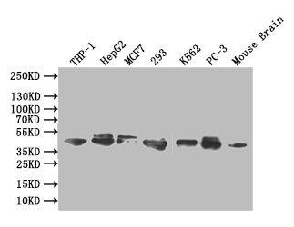
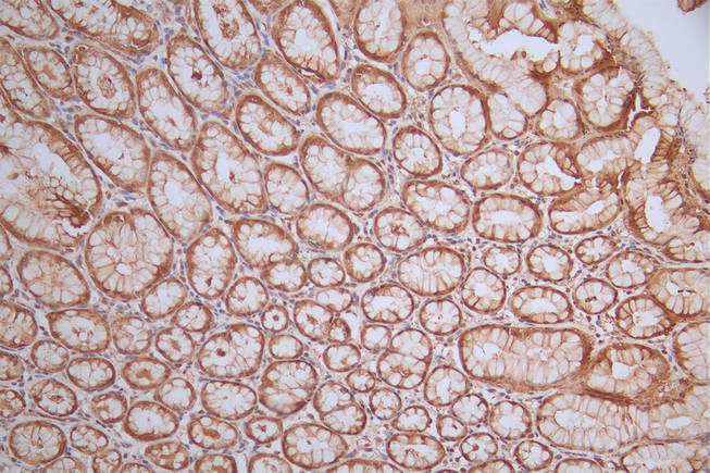
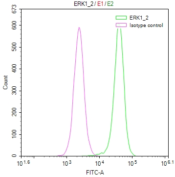
Validation across multiple platforms demonstrates broad experimental utility. Western blot analysis at 1:1000 dilution reveals clean detection of both the 42 kDa ERK2 and 44 kDa ERK1 bands, matching predicted molecular weights, across an extensive panel including THP-1, HepG2, MCF7, HEK293, K562, and PC-3 cell lysates, as well as mouse brain tissue. This cross-species reactivity between human and mouse samples supports translational research workflows. Immunohistochemistry validation in paraffin-embedded human stomach tissue confirms suitability for tissue-based studies, while flow cytometry analysis of HeLa cells demonstrates clear signal separation from isotype controls.
Whether investigating MAPK pathway dysregulation in cancer models, characterizing upstream signaling events, or validating pathway inhibitor efficacy, this antibody provides the technical foundation for confident ERK1/2 detection across western blotting, immunohistochemistry, and flow cytometry applications.
There are currently no reviews for this product.

