MRAS Recombinant Monoclonal Antibody

Code CSB-RA014781MA1HU
Size US$210
Order now
Image
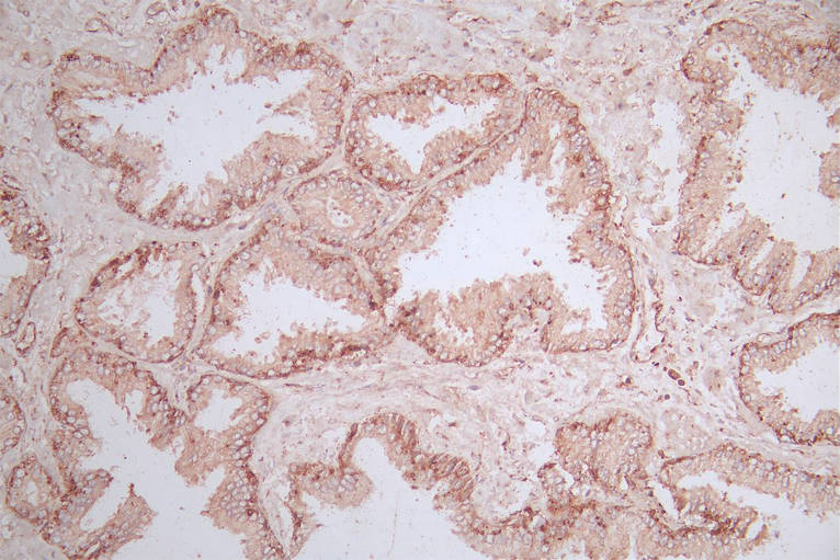
  • IHC image of CSB-RA014781MA1HU diluted at 1:600 and staining in paraffin-embedded human prostate tissue performed on a Leica BondTM system. After dewaxing and hydration, antigen retrieval was mediated by high pressure in a citrate buffer (pH 6.0). Section was blocked with 10% normal goat serum 30min at RT. Then primary antibody (1% BSA) was incubated at 4°C overnight. The primary is detected by a Goat anti-Mouse IgG labeled by HRP and visualized using 0.05% DAB.
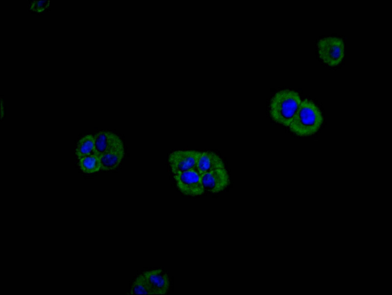
  • Immunofluorescence staining of Hela cell with CSB-RA014781MA1HU at 1:200, counter-stained with DAPI. The cells were fixed in 4% formaldehyde and blocked in 10% normal Goat Serum. The cells were then incubated with the antibody overnight at 4C. The secondary antibody was FITC-conjugated AffiniPure Goat Anti-Mouse IgG(H+L).
Have Questions? Leave a Message or Start an on-line Chat

Product Details

Uniprot No.
Target Names
Alternative Names
FLJ42964 antibody; M ras antibody; M-ras antibody; Mras antibody; Muscle and microspikes Ras antibody; Muscle RAS oncogene homolog antibody; Muscle Ras oncogene homologue antibody; Muscle Ras viral oncogene homolog antibody; R ras3 antibody; R-ras3 antibody; Ras related protein MRas antibody; Ras related protein RRas3 antibody; Ras-related protein M-Ras antibody; Ras-related protein R-Ras3 antibody; RASM_HUMAN antibody; Related Ras viral oncogene homolog 3 antibody; RRas3 antibody; XRas antibody
Species Reactivity
Human
Immunogen
Recombinant Human MRAS protein
Immunogen Species
Homo sapiens (Human)
Conjugate
Non-conjugated
Clonality
Monoclonal
Isotype
Mouse IgG2a
Clone No.
17H12
Purification Method
Affinity-chromatography
Concentration
It differs from different batches. Please contact us to confirm it.
Buffer
Preservative: 0.03% Proclin 300
Constituents: 50% Glycerol, 0.01M PBS, PH 7.4
Form
Liquid
Tested Applications
ELISA, IHC, IF
Recommended Dilution
Application Recommended Dilution
IHC 1:20-1:200
IF 1:20-1:200
Troubleshooting and FAQs
Storage
Upon receipt, store at -20°C or -80°C. Avoid repeated freeze.
Lead Time
Basically, we can dispatch the products out in 1-3 working days after receiving your orders. Delivery time maybe differs from different purchasing way or location, please kindly consult your local distributors for specific delivery time.
Description

MRAS, also known as R-Ras3, is a member of the Ras superfamily of small GTPases that plays a distinctive role in cellular signaling networks. Unlike its more extensively studied relatives, MRAS functions as a modulator of MAP kinase pathways and has been implicated in cell adhesion, migration, and differentiation processes. Its involvement in signal transduction cascades that influence cell fate decisions makes it a compelling target for researchers investigating oncogenic mechanisms and developmental biology.

This recombinant monoclonal antibody, generated from clone 17H12, offers the reproducibility and consistency that demanding experimental workflows require. Because recombinant antibodies are produced from defined sequences rather than hybridoma supernatants, researchers can expect uniform performance across lots, eliminating a common source of experimental variability. The antibody was raised against recombinant human MRAS protein and has been validated for human sample detection.

Experimental validation demonstrates versatility across multiple detection platforms. Immunohistochemistry performed on paraffin-embedded human prostate tissue using a Leica Bond system with citrate buffer antigen retrieval shows specific staining at dilutions ranging from 1:50 to 1:200. Immunofluorescence studies in HeLa cells reveal clear signal when used at 1:200 dilution, with the antibody successfully detecting MRAS in formaldehyde-fixed samples. These validated protocols provide researchers with established starting points for their own investigations.

The affinity-purified IgG2a format and glycerol-stabilized formulation support long-term storage stability at -20°C or -80°C. For investigators exploring cancer biology or signal transduction mechanisms, this antibody provides a reliable tool for examining MRAS expression patterns and localization in human tissues and cell culture models.

Usage
For Research Use Only. Not for use in diagnostic or therapeutic procedures.

Customer Reviews and Q&A

 Customer Reviews

There are currently no reviews for this product.

Submit a Review here

Target Background

Function
Serves as an important signal transducer for a novel upstream stimuli in controlling cell proliferation. Activates the MAP kinase pathway.
Gene References into Functions
  1. acute coronary syndrome in Czechs (30.4% vs 29.4% carriers of the minor T allele of MRAS[recessive model], p = 0.54; OR 1.05; 95% CI 0.89-1.24 for males and 32.1% vs 29.7% carriers of the minor T allele, p = 0.28; OR 1.12; 95% CI 0.91-1.37 for females) PMID: 29264877
  2. Results discovered for the first time that MRAS is recurrently mutated, indicating that MRAS mutations could drive tumorigenesis of Type IV gastric neoplasm. PMID: 27891760
  3. The association of the MARS rs6782181 polymorphism and serum lipid levels is different between the Mulao and Han populations, or between males and females in the both ethnic groups. PMID: 25973078
  4. The MRAS gene loci might have a minor effect in conferring susceptibility to coronary artery disease in the Chinese population. PMID: 25800439
  5. Both MRAS and SHOC2 play a key role in polarized migration. PMID: 24211266
  6. implicated in a novel pathway of neuronal differentiation by coupling specific trophic factors to the MAPK cascade through the activation of B-Raf PMID: 12138204
  7. These findings proved a crucial role of the cross-talk between two Ras-family GTPases M-Ras and Rap1, mediated by RA-GEF-2, in adhesion signaling. PMID: 17538012
  8. Identification of one new CAD risk locus on 3q22.3 in MRAS, and suggestive association with a locus on 12q24.31 near HNF1A-C12orf43. PMID: 19198612

Show More

Hide All

Subcellular Location
Cell membrane; Lipid-anchor; Cytoplasmic side.
Protein Families
Small GTPase superfamily, Ras family
Tissue Specificity
Expression highly restricted to the brain and heart.
Database Links

HGNC: 7227

OMIM: 608435

KEGG: hsa:22808

STRING: 9606.ENSP00000289104

UniGene: Hs.527021

icon of phone
Call us
301-363-4651 (Available 9 a.m. to 5 p.m. CST from Monday to Friday)

icon of address
Address
No. 269, Shendun 5th Road, Donghu Hi-Tech Development Area, Hubei Province, 430206, P.R.China
icon of social media
Join us with

Subscribe newsletter

Leave a message

* To protect against spam, please pass the CAPTCHA test below.
CAPTCHA verification
© 2007-2026 CUSABIO TECHNOLOGY LLC All rights reserved. 鄂ICP备15011166号-1
Place an order now

I. Product details

*
*
*
*

II. Contact details

*
*

III. Ship To

*
*
*
*
*
*
*

IV. Bill To

*
*
*
*
*
*
*
*