Call us
301-363-4651 (Available 9 a.m. to 5 p.m. CST from Monday to Friday)
| Code | CSB-RA943230A0HU |
| Size | US$210 |
| Order now | |
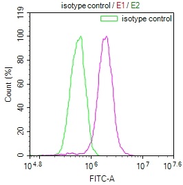
| Image |
|
| Have Questions? | Leave a Message or Start an on-line Chat |
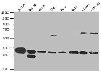
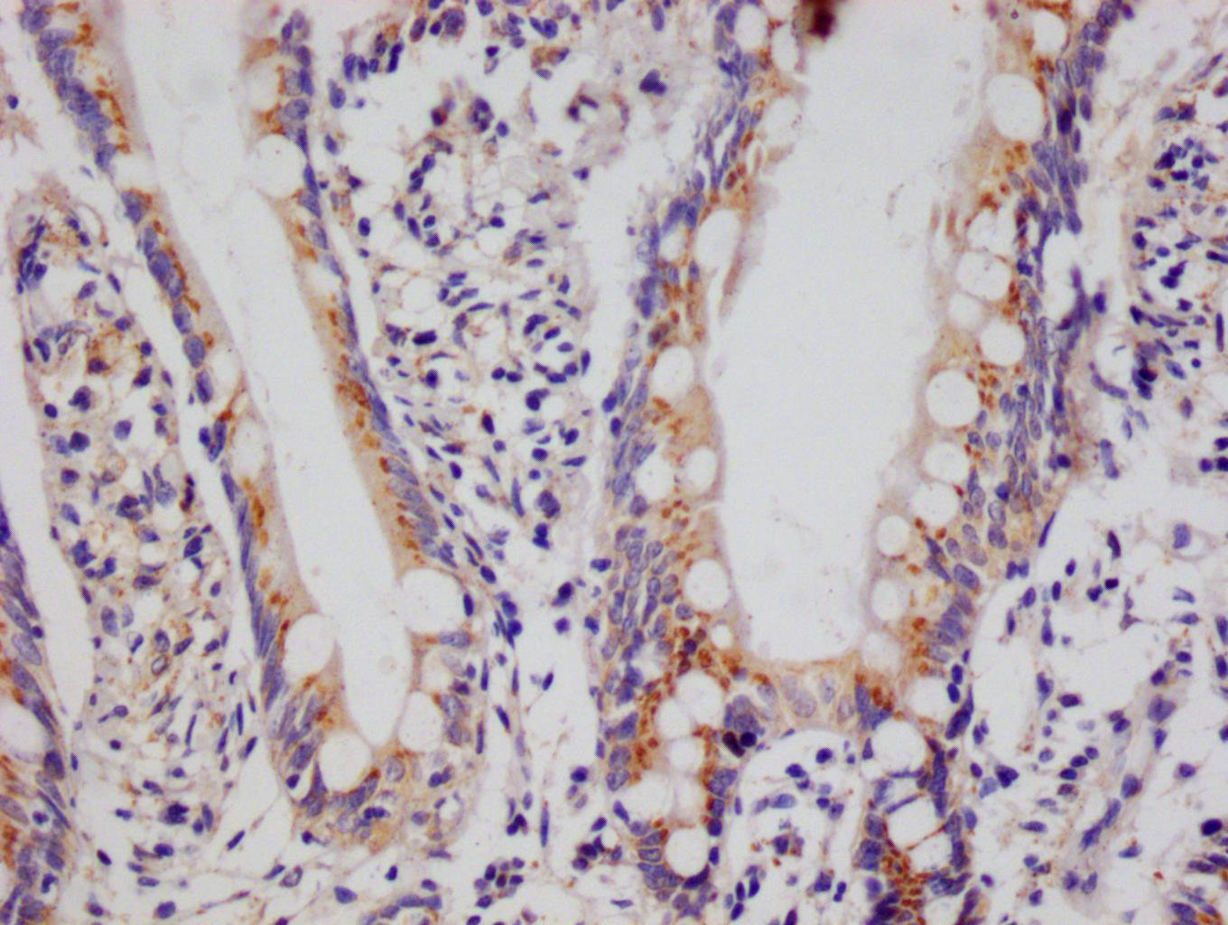
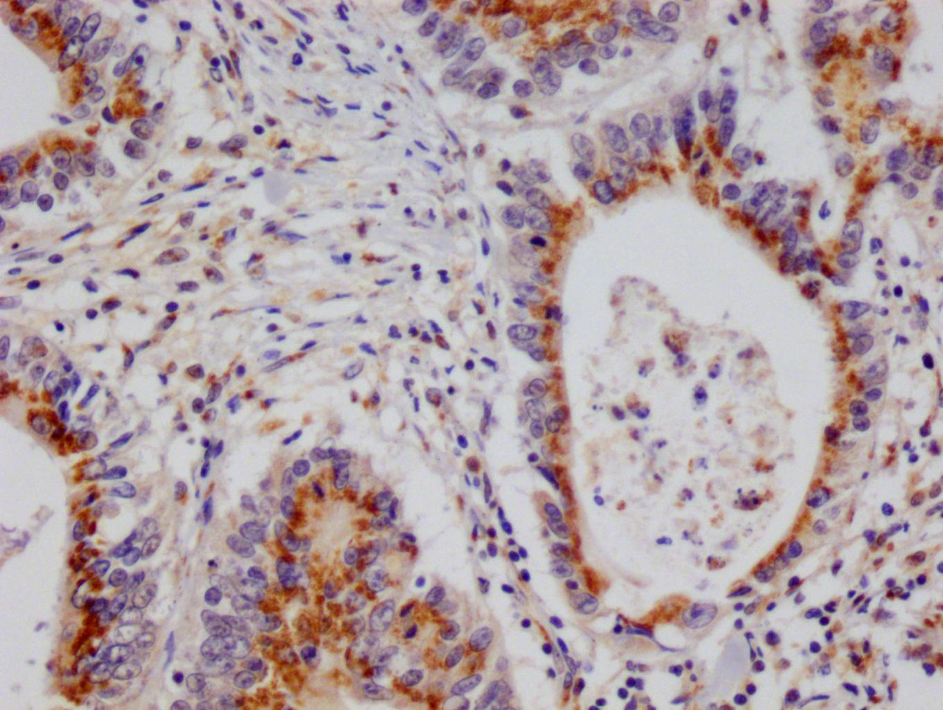
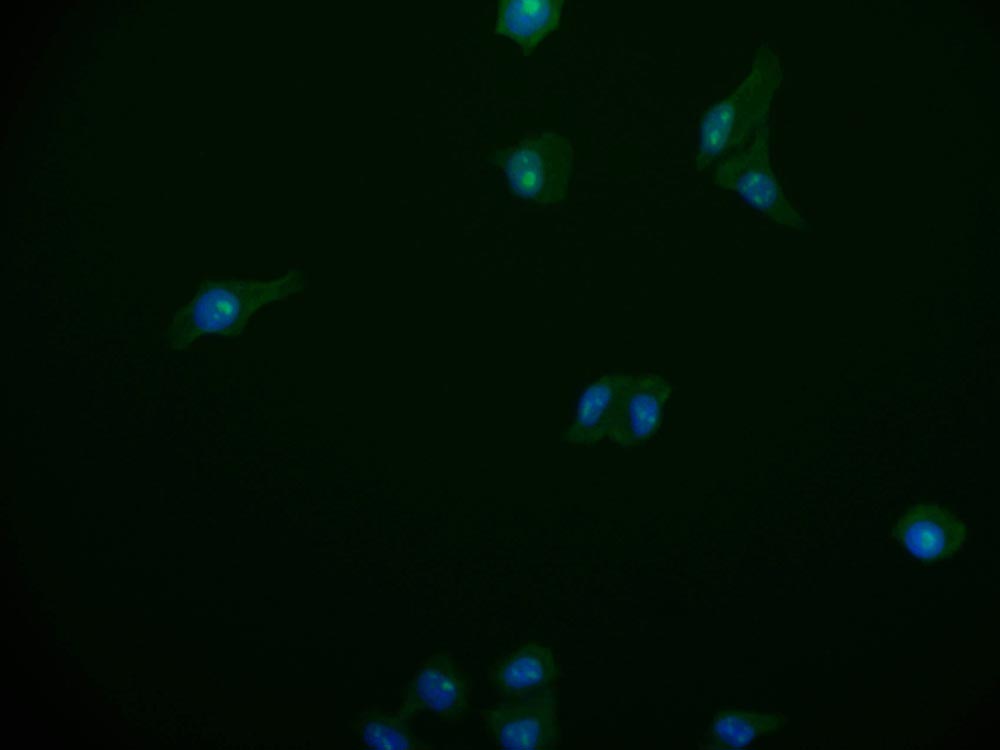
| Application | Recommended Dilution |
|---|---|
| WB | 1:500-1:2000 |
| IHC | 1:50-1:200 |
| IF | 1:50-1:200 |
| FC | 1:50-1:200 |
Generating the PSME1 recombinant monoclonal antibody involves a series of intricate processes. It starts with harvesting the PSME1 monoclonal antibody and sequencing its gene. A vector carrying the PSME1 monoclonal antibody gene is then constructed and introduced into a host cell line for culture. During PSME1 monoclonal antibody production, a synthesized peptide based on human PSME1 is utilized as the immunogen. The PSME1 recombinant monoclonal antibody is subsequently purified using affinity chromatography from the cell culture supernatant and subjected to ELISA, WB, IHC, IF, and FC applications to verify its specificity. It only detects human PSME1 protein.
PSME1, also known as PA28α, is a regulatory subunit of the 20S proteasome complex. It forms a complex with the subunit PSME2 (PA28β) and binds to the ends of the 20S proteasome, activating its proteolytic activity. PSME1 enhances the proteasomal degradation of antigenic proteins presented on MHC class I molecules, promoting their recognition by T lymphocytes. In addition, PSME1 plays a role in cell cycle progression and apoptosis and is involved in the regulation of various cellular processes such as DNA repair and transcriptional regulation.
There are currently no reviews for this product.

