| Image |
-
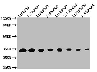
Western Blot
Positive WB detected in: 50ng recombinant protein
All lanes: GFP antibody at 1:50000, 1:100000, 1:200000, 1:400000, 1:800000, 1:1600000, 1:3200000, 1:6400000
Secondary
Goat polyclonal to mouse IgG at 1/50000 dilution
Predicted band size: 32 KDa
Observed band size: 32 KDa
Exposure time:5min
-
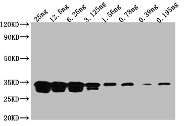
Western Blot
Positive WB detected in: Recombinant protein at 25ng, 12.5ng, 6.25ng, 3.125ng, 1.56ng, 0.78ng, 0.39ng, 0.195ng
All lanes: GFP antibody at 1:2000
Secondary
Goat polyclonal to mouse IgG at 1/50000 dilution
Predicted band size: 32 KDa
Observed band size: 32 KDa
Exposure time:5min
-
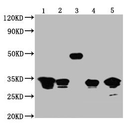
Western Blot
Positive WB detected in: 1-4 lanes:Recombinant proteins with GFP tag for 50ng; 5 lane: 293F whole cell lysate transfected with GFP for 5μg
All lanes GFP antibody at 1:5000
Secondary
Goat polyclonal to mouse IgG at 1/50000 dilution
Predicted band size:1,2,3,4 and 5 is 32,32,50,32,32 KDa respectively
Observed band size: 32,32,50,32,32 KDa
Exposure time:1min
-
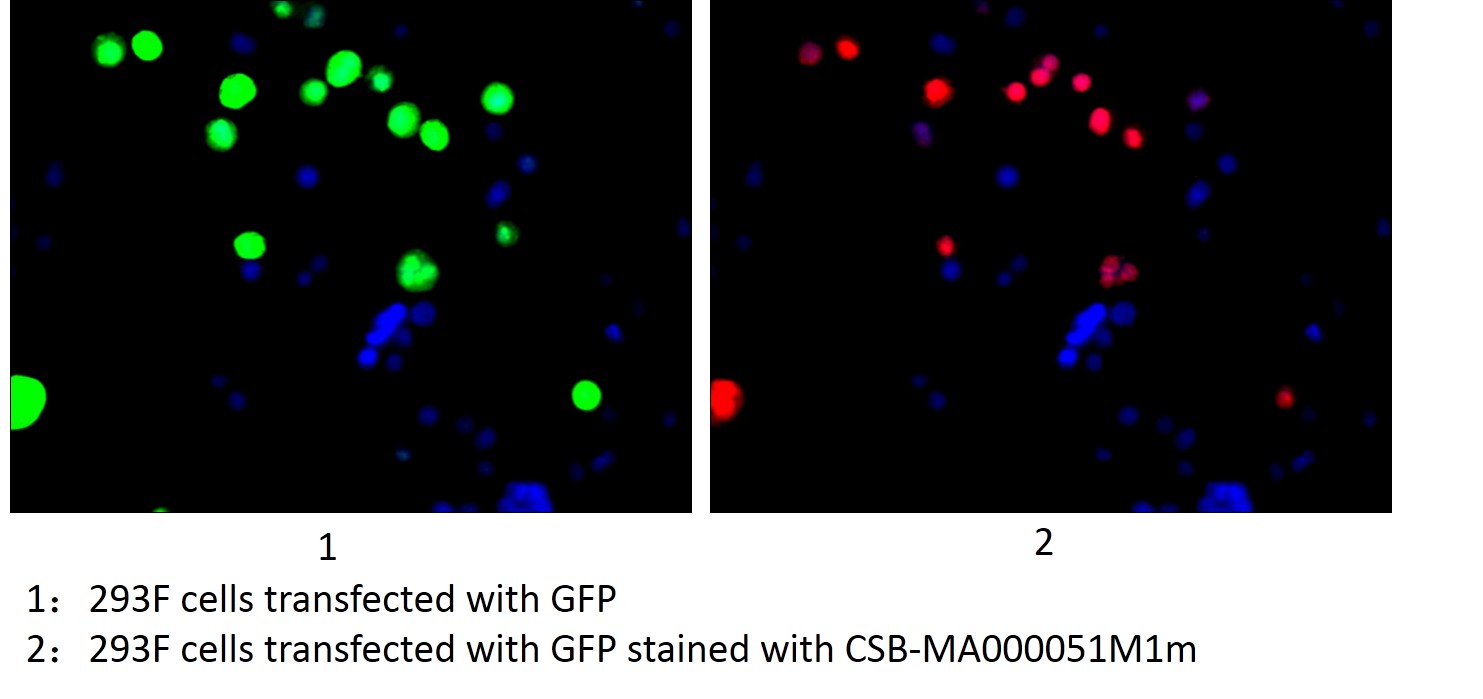
Immunofluorescence staining of 293F cells transfected with GFP with CSB-MA000051M1m at 1:100, counter-stained with DAPI. The cells were fixed in 4% formaldehyde, permeated by 0.2% TritonX-100, and blocked in 10% normal Goat Serum. The cells were then incubated with the antibody overnight at 4°C. Nuclear DNA was labeled in blue with DAPI. The secondary antibody was R-PE-conjugated Goat Anti-Mouse IgG(H+L).
-
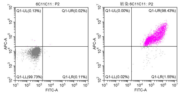
Two-color flow cytometric analysis showing 293F cells untransfected (Left) or transfected with GFP (Right) stained with CSB-MA000051M1m at 1:200. The cells were fixed in 70% ethanol at 4°C overnight. Then 10% normal goat serum was Incubated to block non-specific protein-protein interactions followed by the antibody (1µg/1*106cells) for 1 h at 4°C. The secondary antibody used was Alexa Fluor 647 AffiniPure Donkey Anti-Mouse IgG (H+L) at 1/250 dilution for 30min at 4°C.
-
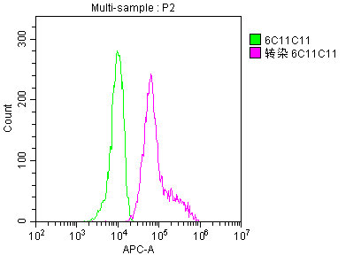
Overlay histogram showing 293F cells transfected with GFP stained with CSB-MA000051M1m (red line) at 1:200. The cells were fixed in 70% ethanol at 4°C overnight. Then 10% normal goat serum was Incubated to block non-specific protein-protein interactions followed by the antibody (1µg/1*106cells) for 1 h at 4°C. The secondary antibody used was Alexa Fluor 647 AffiniPure Donkey Anti-Mouse IgG (H+L) at 1/250 dilution for 30min at 4°C. Isotype control antibody (green line) was mouse IgG2b (1µg/1*106cells) used under the same conditions. Acquisition of >10,000 events was performed.
-
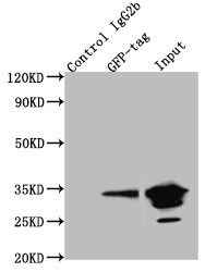
Immunoprecipitating GFP in 293F whole cell lysate transfected with GFP
Lane 1: Mouse control IgG2b instead of CSB-MA000051M1m in 293F whole cell lysate transfected with GFP
Lane 2: CSB-MA000051M1m (4µg) + 293F whole cell lysate transfected with GFP (500µg)
Lane 3: 293F whole cell lysate transfected with GFP (5µg)
For western blotting, the blot was detected with CSB-MA000051M1m at 1:2000, and a HRP-conjugated Protein G antibody was used as the secondary antibody at 1:50000
|