S Antibody

Code CSB-PA336163EA01HIY
Size US$166
Order now
The Latest Promotion Free Antibody trial simple
Have Questions? Leave a Message or Start an on-line Chat

Product Details

Full Product Name
Rabbit anti-Human coronavirus OC43 S Polyclonal antibody
Uniprot No.
Target Names
S
Alternative Names
S antibody; 3Spike glycoprotein antibody; S glycoprotein antibody; E2 antibody; Peplomer protein) [Cleaved into: Spike protein S1; Spike protein S2; Spike protein S2'] antibody
Raised in
Rabbit
Species Reactivity
Human coronavirus OC43
Immunogen
Recombinant Human coronavirus OC43 Spike glycoprotein protein (15-344AA)
Immunogen Species
Human coronavirus OC43
Conjugate
Non-conjugated

The S Antibody (Product code: CSB-PA336163EA01HIY) is Non-conjugated. For S Antibody with conjugates, please check the following table.

Available Conjugates
Conjugate Product Code Product Name Application
HRP CSB-PA336163EB01HIY S Antibody, HRP conjugated ELISA
FITC CSB-PA336163EC01HIY S Antibody, FITC conjugated
Biotin CSB-PA336163ED01HIY S Antibody, Biotin conjugated ELISA
Clonality
Polyclonal
Isotype
IgG
Purification Method
>95%, Protein G purified
Concentration
It differs from different batches. Please contact us to confirm it.
Buffer
Preservative: 0.03% Proclin 300
Constituents: 50% Glycerol, 0.01M PBS, PH 7.4
Form
Liquid
Tested Applications
ELISA
Protocols
Troubleshooting and FAQs
Storage
Upon receipt, store at -20°C or -80°C. Avoid repeated freeze.
Lead Time
Basically, we can dispatch the products out in 1-3 working days after receiving your orders. Delivery time maybe differs from different purchasing way or location, please kindly consult your local distributors for specific delivery time.
Description

Rabbits were immunized with recombinant human coronavirus OC43 spike glycoprotein protein (15-344aa) to generate the CSB-PA321224LA01BUA polyclonal antibody against the spike protein. The spike protein of human coronavirus OC43 is an essential component of the virus and plays a crucial role in its pathogenesis, making it a potential target for the development of vaccines and therapeutics.

Following protein G purification, the rabbit anti-human coronavirus OC43 S polyclonal antibody achieved a purity level of 95%. This non-conjugated IgG specifically binds to the spike protein of human coronavirus OC43 and can detect endogenous levels of the protein, as demonstrated by ELISA.

Customer Reviews and Q&A

 Customer Reviews
Average Rating:
5.0 - 3 reviews

Submit a Review here

Applications : Western Blot Assay

Sample type: Cells

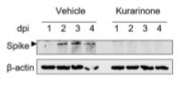
Review: Western blot of the lysates of HCoV-OC43-infected MRC-5 cell streated with kurarinone or vehicle and evaluated at 1,2,3, and 4dpi. The HCoV-OC43 Spike protein was detected and indicated by an arrow head as shown; β-actin was used as a loading control.

By Anonymous

Applications : Immunofluorescence experiments

Sample type: cells

Review: To assess whether 5 min of various UVC illumination reduces the number of infected cells, immunostaining was performed to detect the presence of OC43 viral particles in the host human cells.

By Anonymous

Applications : Western Blot Analysis

Sample type: cell

Review: In MRC-5 and A549 cells was detected using Western blot analysis following HCoV-OC43 infection and vehicle or 10 μM CDN treatment at 1–4 dpi. β-actin was used as the internal control.

By Anonymous

Target Background

Function
S1 attaches the virion to the cell membrane by interacting with sialic acid-containing cell receptors, initiating the infection.; attaches the virion to the cell membrane by interacting with host receptor, initiating the infection.; mediates fusion of the virion and cellular membranes by acting as a class I viral fusion protein. Under the current model, the protein has at least three conformational states: pre-fusion native state, pre-hairpin intermediate state, and post-fusion hairpin state. During viral and target cell membrane fusion, the coiled coil regions (heptad repeats) assume a trimer-of-hairpins structure, positioning the fusion peptide in close proximity to the C-terminal region of the ectodomain. The formation of this structure appears to drive apposition and subsequent fusion of viral and target cell membranes.; Acts as a viral fusion peptide which is unmasked following S2 cleavage occurring upon virus endocytosis.
Subcellular Location
Virion membrane; Single-pass type I membrane protein. Host endoplasmic reticulum-Golgi intermediate compartment membrane; Single-pass type I membrane protein. Host cell membrane; Single-pass type I membrane protein.
Protein Families
Betacoronaviruses spike protein family
CUSABIO guaranteed quality
icon of phone
Call us
301-363-4651 (Available 9 a.m. to 5 p.m. CST from Monday to Friday)
icon of address
Address
7505 Fannin St., Ste 610, Room 7 (CUBIO Innovation Center), Houston, TX 77054, USA
icon of social media
Join us with

Subscribe newsletter

Leave a message

* To protect against spam, please pass the CAPTCHA test below.
CAPTCHA verification
© 2007-2024 CUSABIO TECHNOLOGY LLC All rights reserved. 鄂ICP备15011166号-1
Place an order now

I. Product details

*
*
*
*

II. Contact details

*
*

III. Ship To

*
*
*
*
*
*
*

IV. Bill To

*
*
*
*
*
*
*
*