Full Product Name
Mouse anti-Homo sapiens (Human) PRDX3 Monoclonal antibody
Alternative Names
Antioxidant protein 1 antibody; AOP 1 antibody; AOP-1 antibody; AOP1 antibody; HBC189 antibody; MER5 antibody; MGC104387 antibody; MGC24293 antibody; mitochondrial antibody; peroxiredoxin 3 antibody; Peroxiredoxin III antibody; Peroxiredoxin-3 antibody; PRDX3 antibody; PRDX3_HUMAN antibody; PRO1748 antibody; Protein MER5 homolog antibody; PRX III antibody; Prx-III antibody; PRX3 antibody; SP 22 antibody; SP-22 antibody; SP22 antibody; Thioredoxin dependent peroxide reductase mitochondrial antibody; Thioredoxin-dependent peroxide reductase antibody
Immunogen
Recombinant Human Thioredoxin-dependent peroxide reductase, mitochondrial protein (63-256AA)
Immunogen Species
Homo sapiens (Human)
Purification Method
>95%, Protein G purified
Concentration
It differs from different batches. Please contact us to confirm it.
Buffer
Preservative: 0.03% Proclin 300
Constituents: 50% Glycerol, 0.01M PBS, PH 7.4
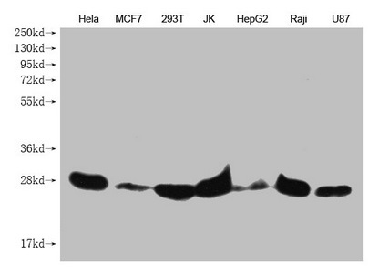
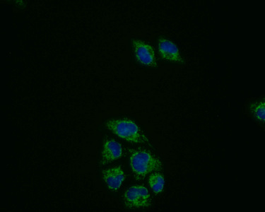
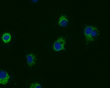
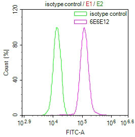
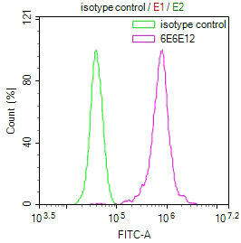
Tested Applications
ELISA, WB, IF, FC
Recommended Dilution
| Application |
Recommended Dilution |
| WB |
WB:1:1000-1:5000 |
| IF |
1:50-1:200 |
| FC |
1:50-1:200 |
Storage
Upon receipt, store at -20°C or -80°C. Avoid repeated freeze.
Lead Time
Basically, we can dispatch the products out in 1-3 working days after receiving your orders. Delivery time maybe differs from different purchasing way or location, please kindly consult your local distributors for specific delivery time.
Usage
For Research Use Only. Not for use in diagnostic or therapeutic procedures.