Call us
301-363-4651 (Available 9 a.m. to 5 p.m. CST from Monday to Friday)
| Code | CSB-MP015007HU |
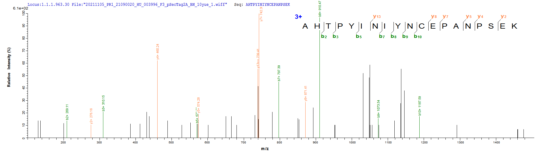
| Abbreviation | Recombinant Human MS4A1 protein-VLPs (Active) |
| MSDS | |
| Size | $558 |
| Order now | |
| Image |
|
| Have Questions? | Leave a Message or Start an on-line Chat |

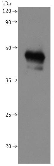
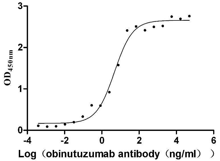
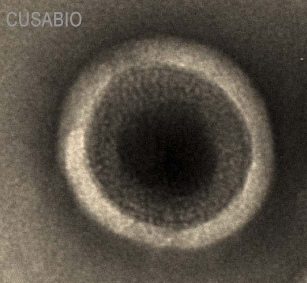
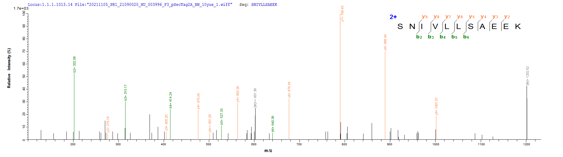
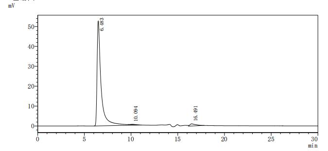
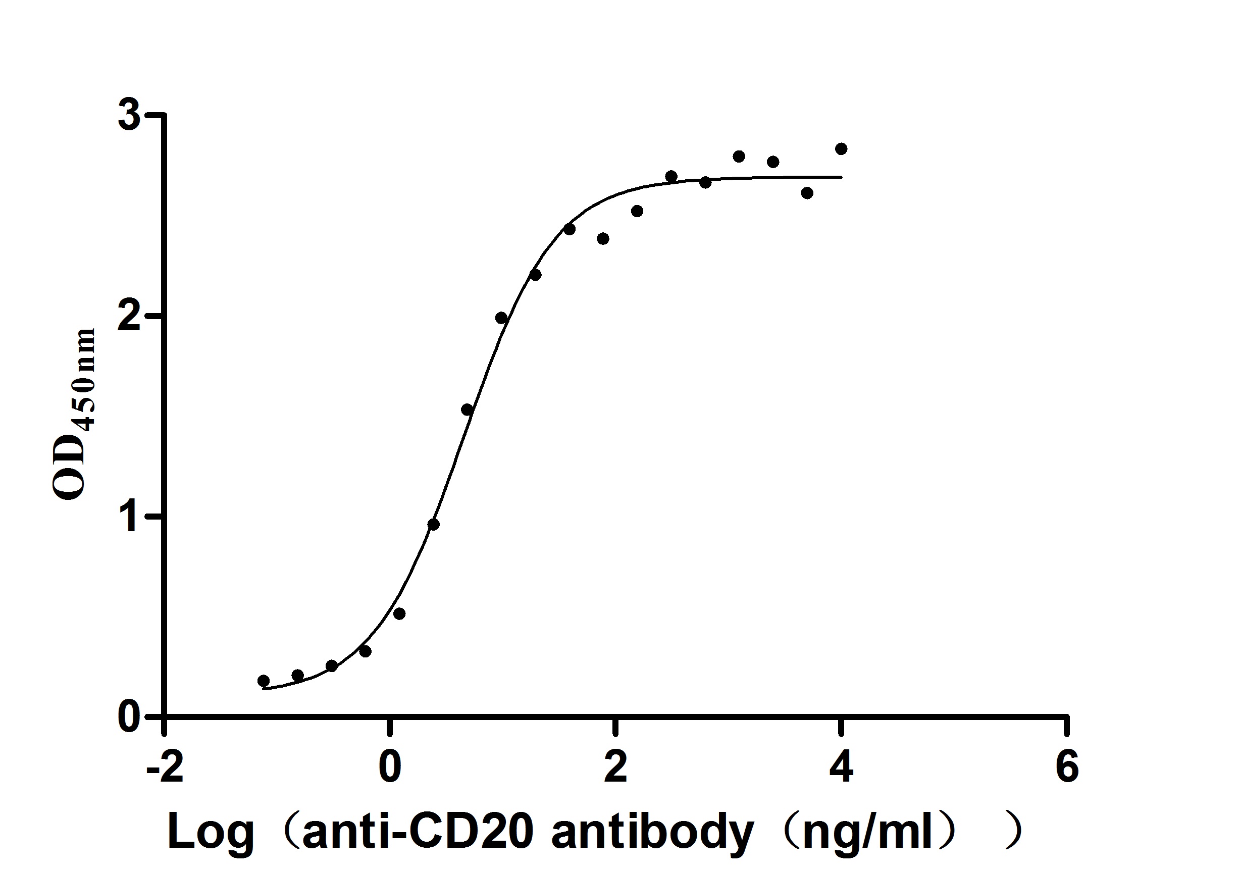
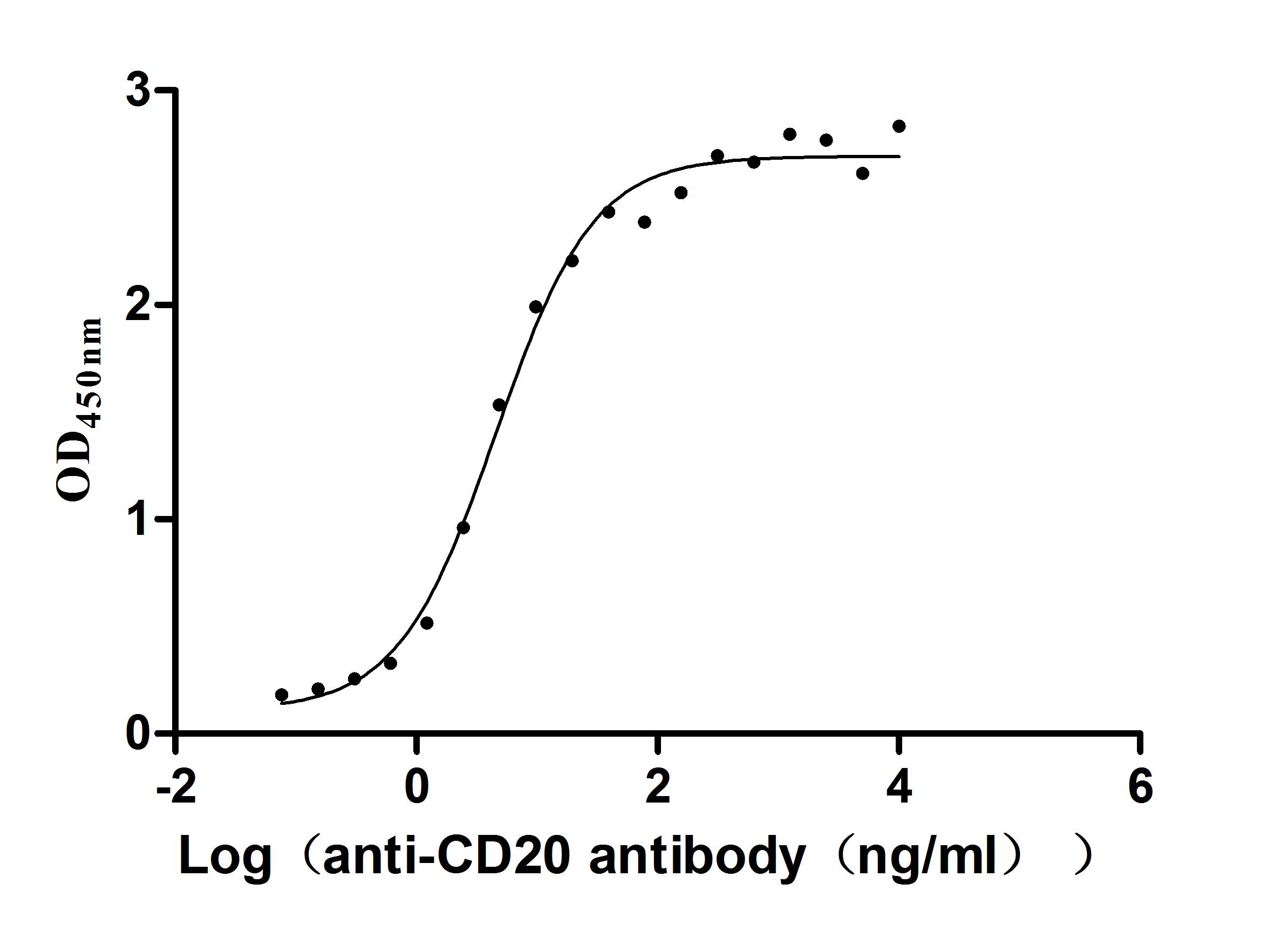
CD20 presents a unique challenge as a multi-pass transmembrane protein that loses native conformation when removed from lipid bilayer context, making virus-like particles (VLPs) an essential format for preserving its four-transmembrane topology. This recombinant human CD20 (MS4A1) construct spans the full-length sequence (aa 1–297) displayed on mammalian cell-derived VLPs, maintaining physiologically relevant membrane orientation critical for conformational epitope recognition. Functional ELISA confirms robust binding activity, with immobilized CD20 at 2 μg/mL engaging anti-CD20 recombinant antibody with an EC50 of 3.243–7.085 ng/mL, supporting use in anti-CD20 therapeutic antibody screening, epitope binning assays, and immunogen preparation for generating conformation-sensitive antibodies in oncology research. The C-terminal 10×His tag (detectable under denaturing conditions) enables protein quantification, while endotoxin levels below 1.0 EU/μg satisfy criteria typical for cell-based binding assays and antibody characterization workflows.
Applications : Antigen
Review: I used the CSB-MP015007HU protein for an ELISA experiment, and the EC50 was measured to be 3.628-5.735 ng/ml, indicating good protein activity. The effect of flow cytometric analysis is also very good, with good repeatability and stable results. Product procurement is convenient and cost-effective.
By Anonymous
Applications : Binding assay/Protein-protein interaction
Review: After receiving the product, we conducted an ELISA experiment with the anti-CD20 (MS4A1) antibody, and the EC50 was 3.628-5.735 ng/ml, showing good binding activity. CD20 (MS4A1) has four times transmembrane domains, I have contacted many companies, but they can\'t express it successfully. CUSABIO expresses full-length proteins and has complete biological activity, which is very powerful!
By Anonymous
Why does the CD20 protein is almost invisible in the tube?
All CUSABIO proteins don’t contain carrier protein or other additives, such as bovine serum albumin (BSA), human serum albumin (HSA), and sucrose. When freeze-drying the lowest salt content solution, it often does not form a white grid structure, but a trace amount of protein deposits within the tube, forming a thin transparent or invisible protein layer.
Tips: Before opening the lid, we recommend centrifuging the tube in a small centrifuge for 20-30 seconds to aggregate the protein attached to the inside wall or cap of the tube to the bottom of the tube. Our quality control steps ensure that the amount of protein contained in each tube is accurate. Although sometimes you can’t see the protein powder, the protein content in the tube is still very accurate.
Does this CD20 protein soluble?
Yes, it is. We offer five expression systems: E. coli, Yeast, Baculovirus, Mammalian cell, and Cell-Free (in vitro E.coli). Among them, yeast only has supernatant expression, and the protein expressed in the supernatant must be a soluble protein.
The E. coli, Baculovirus, Mammalian cell, and Cell-Free (in vitro E.coli) have both supernatant expression and inclusion body expression. As long as the protein is expressed in the supernatant, it must be soluble. If it is expressed in the inclusion body, we will also take a variety of methods to refold it and finally ensure that all the proteins we provide are soluble.
Why should we add the protective agent to the protein solution before lyophilization? What's the general protective agent? Which kind of protective agent do you usually add?
The protective agent is used in the process of freeze-drying and storage to protect the protein.
Commonly used protective agents or stabilizers include saccharides, polyols, polymers, surfactants, some proteins, and amino acids, etc. We usually add 6% (mass ratio by volume) of trehalose as lyoprotectant. Trehalose can significantly prevent the alter of the protein secondary structure and the extension and aggregation of proteins during freeze-drying process. Mannitol is also a universal applied lyoprotectant and filler, which can reduce the aggregation of certain proteins after lyophilization.
Tips: For the majority of proteins, they can be only stored at 4 ℃ for a short term (about a week) after being resuspended. If you want long-term preservation, they should be formulated as a diluent (which must contain a carrier protein, such as 0.1% BSA, 5% HSA, or 10% FBS), and then repackaged and frozen at -20℃ or -80℃. Be sure to avoid repeated freezing and thawing. Because each freezing and thawing cycle will cause part of the protein inactivation.
Is the CD20 protein free of animal components?
Yes, it is. We guarantee that all proteins produced by CUSABIO are 100% animal-free, as we do not use any animal components in our raw materials and no animal components are added during the production process. Serum-free media is also used in the expression of our mammalian proteins. We can provide a declaration of no animal component if it is required.
Is this protein cell-component-free?
The protein expressed in the body, regardless of the system, is expressed by cell, and the cell expression can be divided into intracellular expression and secretory expression.
The E. coli system only has intracellular expression and needs to be broken, so the cell components are relatively more secreted than other systems.
Yeast, Baculovirus, and Mammalian cell systems can be expressed both in the cell and in the secretory expression, and the secreted expression of the protein component remains relatively less intracellular.
In vitro expression means that cell-free and the E. coli cell extract is also added, so that the cell component remains.
Therefore, no matter which expression system and which way the protein is expressed, there will be residual cell components, but we finally use affinity chromatography to purify. In theory, the residual of the cell components will be very small, but 100% of no residue cannot be guaranteed. (Nor does any companies dare to guarantee).
Can you offer aseptic manufacture processing?
Yes, we can offer an aseptic processing service and it is free of charge, but you should remark this information when placing the order. We've performed aseptic processing for liquid protein before lyophilization, but there may exist contamination during lyophilization process. To clarify, lyophilized proteins can’t guarantee aseptic processing for the whole manufacture process.
Can you remove the tag?
Not all protein tags can be removed as some proteins will be very unstable after tag removal. Please contact us in advance if you need to remove the tag which takes 2-3 business days. If we succeed in removing the tag, we will charge for extra cost for it. If we fail in removing the tag, we won't charge for any extra cost and remark this information in the datasheet as follows "Note: The laboratory determined that the tag on your protein could not be removed with standard laboratory procedures. Your protein is being supplied with the tag intact."
What is the concentration range of the protein? How do you determine the quantity of it?
The protein concentration of each batch won't be exactly the same but we could guarantee 0.1-5.0 mg/mL concentration for our expression system. If you have a special requirement for protein concentration, please contact us in advance.
There are three methods of protein concentration detection: bradford method, BCA method, A280 method. Each of these three methods has interference buffer components. According to the composition of buffer, the bradford method is the most popular with laboratories including ours, and the concentration detection range is 0.1-5 mg/mL. We also use BCA method in times.

