RD3 Antibody

Code CSB-PA800101LA01HU
Size US$166
Order now
Image
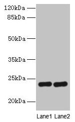
  • Western blot
    All lanes: RD3 antibody at 0.5µg/ml
    Lane 1: Mouse thymus tissue
    Lane 2: Human placenta tissue
    Secondary
    Goat polyclonal to rabbit IgG at 1/10000 dilution
    Predicted band size: 23 kDa
    Observed band size: 23 kDa

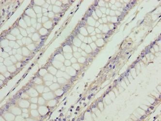
  • Immunohistochemistry of paraffin-embedded human colon cancer using CSB-PA800101LA01HU at dilution of 1:100

Have Questions? Leave a Message or Start an on-line Chat

Product Details

Full Product Name
Rabbit anti-Homo sapiens (Human) RD3 Polyclonal antibody
Uniprot No.
Target Names
RD3
Alternative Names
RD3 antibody; C1orf36 antibody; Protein RD3 antibody; Retinal degeneration protein 3 antibody
Raised in
Rabbit
Species Reactivity
Human, Mouse
Immunogen
Recombinant Human Protein RD3 protein (1-195AA)
Immunogen Species
Homo sapiens (Human)
Conjugate
Non-conjugated

The RD3 Antibody (Product code: CSB-PA800101LA01HU) is Non-conjugated. For RD3 Antibody with conjugates, please check the following table.

Available Conjugates
Conjugate Product Code Product Name Application
HRP CSB-PA800101LB01HU RD3 Antibody, HRP conjugated ELISA
FITC CSB-PA800101LC01HU RD3 Antibody, FITC conjugated
Biotin CSB-PA800101LD01HU RD3 Antibody, Biotin conjugated ELISA
Clonality
Polyclonal
Isotype
IgG
Purification Method
Antigen Affinity Purified
Concentration
It differs from different batches. Please contact us to confirm it.
Buffer
Preservative: 0.03% Proclin 300
Constituents: 50% Glycerol, 0.01M PBS, PH 7.4
Form
Liquid
Tested Applications
ELISA, WB, IHC
Recommended Dilution
Application Recommended Dilution
WB 1:1000-1:5000
IHC 1:20-1:200
Troubleshooting and FAQs
Storage
Upon receipt, store at -20°C or -80°C. Avoid repeated freeze.
Lead Time
Basically, we can dispatch the products out in 1-3 working days after receiving your orders. Delivery time maybe differs from different purchasing way or location, please kindly consult your local distributors for specific delivery time.
Usage
For Research Use Only. Not for use in diagnostic or therapeutic procedures.

Customer Reviews and Q&A

 Customer Reviews

There are currently no reviews for this product.

Submit a Review here

Target Background

Function
Plays a critical role in the regulation of enzymes involved in nucleotide cycle in photoreceptors. Inhibits the basal catalytic activity and the GCAP-stimulated activity of GUCY2D and GUCY2F, two retinal guanylyl cyclases involved in the production of cGMP in photoreceptors. Involved in the transport of GUCY2D and GUCY2F to their target sites in the photoreceptor outer segment. Up-regulates the activity of GUK1, a kinase that plays also an essential role for recycling GMP and indirectly, cGMP. Plays an important role for the survival of rods and cones in the retina.
Gene References into Functions
  1. Re-expressing RD3 in metastatic site-derived aggressive cells reverted their metastatic potential both in vitro and in vivo. Conversely muting RD3 in neuroblastoma cells not only heightened invasion/migration but also dictated aggressive disease with metastasis. PMID: 26375249
  2. This study reports the results of an international study aimed at delineating the clinical and molecular spectrum of RD3 mutations in retinal dystrophies. PMID: 23308101
  3. Mutations in RD3 are a very rare cause of Leber's congenital amaurosis (LCA) associated with an extremely severe form of retinal dystrophy. PMID: 22531706
  4. RD3 suppresses the basal catalytic activity of guanylyl cyclase activating proteins (GCAP) in a noncompetitive manner. PMID: 21928830
  5. Identification and sequence analysis of C1orf36. PMID: 12914764
  6. the retinopathy-associated RD3 protein is part of subnuclear protein complexes involved in diverse processes, such as transcription and splicing. PMID: 17186464

Show More

Hide All

Involvement in disease
Leber congenital amaurosis 12 (LCA12)
Subcellular Location
Cell projection, cilium, photoreceptor outer segment. Photoreceptor inner segment. Endosome. Nucleus. Cytoplasm. Cytoplasm, perinuclear region.
Tissue Specificity
Expressed in retina. Widely expressed (at protein level). In the retina the strongest immunoreactivity is detected in the inner half of the cytoplasmic portion of the photoreceptor layer, where rods and cones are found, and the external half of the outer
Database Links

HGNC: 19689

OMIM: 180040

KEGG: hsa:343035

STRING: 9606.ENSP00000355969

UniGene: Hs.632495

icon of phone
Call us
301-363-4651 (Available 9 a.m. to 5 p.m. CST from Monday to Friday)
icon of address
Address
7505 Fannin St., Ste 610, Room 7 (CUBIO Innovation Center), Houston, TX 77054, USA
icon of social media
Join us with

Subscribe newsletter

Leave a message

* To protect against spam, please pass the CAPTCHA test below.
CAPTCHA verification
© 2007-2025 CUSABIO TECHNOLOGY LLC All rights reserved. 鄂ICP备15011166号-1
Place an order now

I. Product details

*
*
*
*

II. Contact details

*
*

III. Ship To

*
*
*
*
*
*
*

IV. Bill To

*
*
*
*
*
*
*
*