RIPK3 Antibody

Code CSB-PA897497ESR1HU
Size US$166
Order now
Image
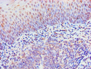
  • Immunohistochemistry of paraffin-embedded human tonsil tissue using CSB-PA897497ESR1HU at dilution of 1:100

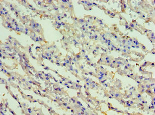
  • Immunohistochemistry of paraffin-embedded human lung tissue using CSB-PA897497ESR1HU at dilution of 1:100

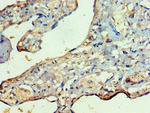
  • Immunohistochemistry of paraffin-embedded human placenta tissue using CSB-PA897497ESR1HU at dilution of 1:100

Have Questions? Leave a Message or Start an on-line Chat

Product Details

Full Product Name
Rabbit anti-Homo sapiens (Human) RIPK3 Polyclonal antibody
Uniprot No.
Target Names
RIPK3
Alternative Names
Receptor interacting protein 3 antibody; Receptor interacting serine threonine kinase 3 antibody; Receptor interacting serine/threonine protein kinase 3 antibody; Receptor-interacting protein 3 antibody; Receptor-interacting serine/threonine-protein kinase 3 antibody; RIP 3 antibody; RIP like protein kinase 3 antibody; RIP-3 antibody; RIP-like protein kinase 3 antibody; RIPK 3 antibody; RIPK3 antibody; RIPK3_HUMAN antibody
Raised in
Rabbit
Species Reactivity
Human
Immunogen
Recombinant Human Receptor-interacting serine/threonine-protein kinase 3 protein (1-120AA)
Immunogen Species
Homo sapiens (Human)
Conjugate
Non-conjugated
Clonality
Polyclonal
Isotype
IgG
Purification Method
Antigen Affinity Purified
Concentration
It differs from different batches. Please contact us to confirm it.
Buffer
PBS with 0.02% sodium azide, 50% glycerol, pH7.3.
Form
Liquid
Tested Applications
ELISA, IHC
Recommended Dilution
Application Recommended Dilution
IHC 1:20-1:200
Troubleshooting and FAQs
Storage
Upon receipt, store at -20°C or -80°C. Avoid repeated freeze.
Lead Time
Basically, we can dispatch the products out in 1-3 working days after receiving your orders. Delivery time maybe differs from different purchasing way or location, please kindly consult your local distributors for specific delivery time.

Customer Reviews and Q&A

 Customer Reviews

There are currently no reviews for this product.

Submit a Review here

Target Background

Function
Serine/threonine-protein kinase that activates necroptosis and apoptosis, two parallel forms of cell death. Necroptosis, a programmed cell death process in response to death-inducing TNF-alpha family members, is triggered by RIPK3 following activation by ZBP1. Activated RIPK3 forms a necrosis-inducing complex and mediates phosphorylation of MLKL, promoting MLKL localization to the plasma membrane and execution of programmed necrosis characterized by calcium influx and plasma membrane damage. In addition to TNF-induced necroptosis, necroptosis can also take place in the nucleus in response to orthomyxoviruses infection: following ZBP1 activation, which senses double-stranded Z-RNA structures, nuclear RIPK3 catalyzes phosphorylation and activation of MLKL, promoting disruption of the nuclear envelope and leakage of cellular DNA into the cytosol. Also regulates apoptosis: apoptosis depends on RIPK1, FADD and CASP8, and is independent of MLKL and RIPK3 kinase activity. Phosphorylates RIPK1: RIPK1 and RIPK3 undergo reciprocal auto- and trans-phosphorylation. In some cell types, also able to restrict viral replication by promoting cell death-independent responses. In response to Zika virus infection in neurons, promotes a cell death-independent pathway that restricts viral replication: together with ZBP1, promotes a death-independent transcriptional program that modifies the cellular metabolism via up-regulation expression of the enzyme ACOD1/IRG1 and production of the metabolite itaconate. Itaconate inhibits the activity of succinate dehydrogenase, generating a metabolic state in neurons that suppresses replication of viral genomes. RIPK3 binds to and enhances the activity of three metabolic enzymes: GLUL, GLUD1, and PYGL. These metabolic enzymes may eventually stimulate the tricarboxylic acid cycle and oxidative phosphorylation, which could result in enhanced ROS production.; (Microbial infection) In case of herpes simplex virus 1/HHV-1 infection, forms heteromeric amyloid structures with HHV-1 protein RIR1/ICP6 which may inhibit RIPK3-mediated necroptosis, thereby preventing host cell death pathway and allowing viral evasion.
Gene References into Functions
  1. The major function of RIP1 kinase activity in TNF-induced necroptosis is to autophosphorylate serine 161. This specific phosphorylation then enables RIP1 to recruit RIP3 and form a functional necrosome, a central controller of necroptosis. PMID: 28176780
  2. RIPK3-dependent cell death and inflammasome activation in FLT3-internal-tandem-duplication-expressing leukemia-initiating cells PMID: 27517160
  3. The necroptosis-inducing kinase RIPK3 reduces adipose tissue inflammation and glucose intolerance. PMID: 27323669
  4. We showed that RIP3 spontaneously drives a necroptosis-induced inflammation in established intestinal cell lines and in ileal/colonic samples from IBD patients. PMID: 28844856
  5. These data demonstrate that caspase-8 functions in synovial antigen-presenting cells to regulate the response to inflammatory stimuli by controlling RIPK3 action, and this delicate balance maintains homeostasis within the joint. PMID: 28978351
  6. The induced expression of RIP3 by UHRF1 RNAi depends on the presence of Sp1. Remarkably, the ectopic expression of RIP3 in RIP3-null cancer cells results in a decrease in tumor growth in mice. Therefore, our findings offer insights into RIP3 expression control in cancer cells and suggest an inhibitory effect of RIP3 on tumorigenesis. PMID: 28981102
  7. 2-hydroxyglutarate bound to DNMT1 and stimulated its association with the RIP3 promoter, inducing hypermethylation that reduces RIP3 protein and consequently impaired RIP3-dependent necroptosis. PMID: 28564603
  8. the in vivo effects were diametrically reversed with RIP3 deletion or RIP1 blockade, resulting in marked tumor protection. The dichotomy between the in vivo and in vitro results suggests that the microenvironmental milieu resulting from RIP1/RIP3 signaling is likely responsible for its protumorigenic effects PMID: 27932417
  9. Shikonin induces glioma cell necroptosis in vitro by reactive oxygen species overproduction and promoting RIP1/RIP3 necrosome formation. PMID: 28816233
  10. In critically ill trauma patients, plasma levels of the necroptosis mediator RIP3 at 48 h were associated with AKI stage and RBC transfusions. PMID: 26925809
  11. our results reveal that the necroptosis adaptor RIPK3 has key anti-inflammatory and anti-tumoral functions in the intestine, and define RIPK3 as a novel colon tumor suppressor PMID: 27344176
  12. adhesion-induced eosinophil cytolysis takes place through RIPK3-MLKL-dependent necroptosis, which can be counterregulated by autophagy PMID: 28412393
  13. Renal clear cell carcinoma cells cells express increased amounts of RIPK1 and RIPK3 and are poised to undergo necroptosis in response to TNFR1 signaling. PMID: 27362805
  14. The results highlight a new role of TSC2 in protecting glioblastoma against photodynamic therapy-induced cell death, and TSC2 and YWHAZ as new RIP3 partners. PMID: 27984090
  15. inactivation of RIP1/RIP3 resulted in reduction of SOCS1 protein levels and partial differentiation of AML cells. AML cells with inactivated RIP1/RIP3 signaling show increased sensitivity to IFN-gamma-induced differentiation. PMID: 27748372
  16. results reveal a pathway for MLKL-dependent programmed necrosis that is executed in the absence of RIPK3 and potentially drives the pathogenesis of severe liver diseases. PMID: 27756058
  17. Necroptosis signaling is modulated by the kinase RIPK1 and requires the kinase RIPK3 and the pseudokinase MLKL. (Review) PMID: 26865533
  18. Results demonstrate that RIPK3 restricts malignant myeloproliferation by activating the inflammasome, which promotes differentiation and cell death, and that loss of RIPK3 increases leukemic burden in mice. Reduced RIPK3 expression is observed across several human acute myeloid leukemia subtypes. PMID: 27411587
  19. Data identify RIPK3 and the inflammasome as key tumor suppressors in acute myeloid leukemia (AML). PMID: 27411587
  20. The main route of cell death induced by shikonin is RIP1K-RIP3K-mediated necroptosis. PMID: 26496737
  21. Results suggest that impaired hepatic proteasome function by alcohol exposure may contribute to hepatic accumulation of RIP3 resulting in necroptosis and steatosis while RIP1 kinase activity is important for alcohol-induced inflammation. PMID: 26769846
  22. The expression level of RIP3K was significantly lower in the malignant tumors. PMID: 26749282
  23. CNOT3 suppression promotes necroptosis by stabilizing mRNAs for cell death-inducing proteins, Ripk1 and Ripk3. PMID: 26437789
  24. RIPK3 expression may allow unmasking the necroptotic signalling machinery in melanoma and points to reactivation of this pathway as a treatment option for metastatic melanoma. PMID: 26355347
  25. Additionally, later in infection, RIP3 is cleaved by the coxsackievirus B3-encoded cysteine protease 3C(pro), which serves to abrogate RIP3-mediated necrotic signaling and induce a nonnecrotic form of cell death. PMID: 26269957
  26. although JNK activation and RIP3 expression are induced by FS, neither contributes to the liver injury. PMID: 25423287
  27. The RIP3 expression is reduced in tumors compared to normal tissue in 85% of breast cancer patients, suggesting that RIP3 deficiency is positively selected during tumor growth/development. PMID: 25952668
  28. Data implicate the infiltrating macrophages as a source of damaging inflammasomes after photoreceptor detachment in a RIP3-dependent manner and suggest a novel therapeutic target for treatment of retinal diseases. PMID: 25906154
  29. PolyIC stimulation of cervical cancer cells induced necroptotic cell death, which was strictly dependent on the expression of the receptor-interacting protein kinase RIPK3. PMID: 25888634
  30. Data suggest that neoalbaconol-induced necroptosis include receptor interacting serine/threonine kinase 1-dependent expression of tumor necrosis factor alpha and receptor interacting serine/threonine kinase 3-dependent generation of reactive oxygen species. PMID: 25575821
  31. Herpes simplex virus 1- and 2 ICP6 and ICP10 proteins prevent necroptosis in human cells by inhibiting the interaction between receptor-interacting protein kinase 1 (RIP1) and RIP3, a key step in tumor necrosis factor (TNF)-induced necroptosis. PMID: 25674983
  32. High expression of RIP3 in keratinocytes from toxic epidermal necrolysis patients potentiates MLKL phosphorylation/activation and necrotic cell death. PMID: 25748555
  33. RIP3 silencing in leukemia cells results in suppression of the complex regulation of the apoptosis/necroptosis switch and NF-kappaB activity. PMID: 25144719
  34. Suppression of RIP3-dependent necroptosis by human cytomegalovirus PMID: 25778401
  35. RIP3 activation following the induction of necroptosis requires the activity of an HSP90 and CDC37 cochaperone complex. PMID: 25852146
  36. The results of this study showed that cerebral ischemia activates transcriptional changes that lead to an increase in the endogenous RIP3 protein level. PMID: 24746856
  37. Enhanced RIP3 signaling in aneurysmal tissues contributes to abdominal aortic aneursym progression by causing smooth muscle cell necroptosis, as well as stimulating vascular inflammation. PMID: 25563840
  38. RIP3-dependent necroptosis mediates non-alcoholic steatohepatitis-induced liver fibrosis via activation of JNK, MCP-1-mediated recruitment of monocytes, and an expansion of intrahepatic biliary/progenitor cells. PMID: 24963148
  39. RIPK3 serves as a negative regulator of selective autophagy by regulating regulates p62-LC3 complex formation via the caspase-8-dependent cleavage of p62 PMID: 25450619
  40. RIP3 holds both necroptosis and apoptosis in balance through a Ripoptosome-like platform. PMID: 25459880
  41. an alanine residue substitution for Ser(89) enhanced RIP1 kinase activity and TNF-induced programmed necrosis without affecting RIP1-RIP3 necrosome formation. PMID: 24059293
  42. targeting the RIP kinase signalling pathway could be an effective therapeutic intervention in retinal degeneration patients. PMID: 24413151
  43. RIP3-mediated MLKL phosphorylation, though important for downstream signaling, is dispensable for stable complex formation between RIP3 and MLKL. PMID: 24095729
  44. Report role of MLKL/RIP3 pathway in necrotic membrane disruption. PMID: 24703947
  45. RIP3 protein expression is significantly increased in the inflamed tissue of inflammatory boowel disease pediatric patients. PMID: 24322838
  46. The protein levels of crucial modulators of necroptosis, RIP1 and RIP3, are increased by shikonin treatment in primary tumor tissues. PMID: 24314238
  47. the importance of the RIP3-MLKL interaction in the formation of functional necrosomes and suggest that translocation of necrosomes to mitochondria-associated membranes is essential for necroptosis signaling. PMID: 23612963
  48. procaspase-8 activity is essential for cell survival by inhibiting both apoptotic and nonapoptotic cell death dependent on receptor-interacting protein kinase 1 (RIP1) and RIP3 PMID: 23071110
  49. Study shows that RIP1 and RIP3 form an amyloid structure through their RIP homotypic interaction motifs and that this heterodimeric amyloid structure is a functional signaling complex that mediates programmed necrosis. PMID: 22817896
  50. study suggests that MLKL is a key RIP3 downstream component of TNF-induced necrotic cell death PMID: 22421439

Show More

Hide All

Subcellular Location
Cytoplasm, cytosol. Nucleus.
Protein Families
Protein kinase superfamily, TKL Ser/Thr protein kinase family
Tissue Specificity
Highly expressed in the pancreas. Detected at lower levels in heart, placenta, lung and kidney.; [Isoform 3]: Expression is significantly increased in colon and lung cancers.
Database Links

HGNC: 10021

OMIM: 605817

KEGG: hsa:11035

STRING: 9606.ENSP00000216274

UniGene: Hs.268551

icon of phone
Call us
301-363-4651 (Available 9 a.m. to 5 p.m. CST from Monday to Friday)
icon of address
Address
7505 Fannin St., Ste 610, Room 7 (CUBIO Innovation Center), Houston, TX 77054, USA
icon of social media
Join us with

Subscribe newsletter

Leave a message

* To protect against spam, please pass the CAPTCHA test below.
CAPTCHA verification
© 2007-2024 CUSABIO TECHNOLOGY LLC All rights reserved. 鄂ICP备15011166号-1
webinars: DT3C facilitates antibody internalization X
Place an order now

I. Product details

*
*
*
*

II. Contact details

*
*

III. Ship To

*
*
*
*
*
*
*

IV. Bill To

*
*
*
*
*
*
*
*