Call us
301-363-4651 (Available 9 a.m. to 5 p.m. CST from Monday to Friday)
| Code | CSB-EP005346HU |
| Abbreviation | Recombinant Human CHI3L1 protein |
| MSDS | |
| Size | $224 |
| Order now | |
| Image |
|
| Have Questions? | Leave a Message or Start an on-line Chat |
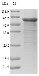
Producing recombinant human chitinase-3-like protein 1 (CHI3L1) starts with isolating the target gene corresponding to the 22-383aa of human CHI3L1. This gene is co-cloned into an expression vector with an N-terminal 10xHis-SUMO-tag and C-terminal Myc-tag gene and introduced into E.coli cells via transformation. The E.coli cells express the protein, which is subsequently harvested from the cell lysate. Purification of the recombinant CHI3L1 protein is commonly achieved using affinity chromatography. The SDS-PAGE is used to assess the purity of this protein, exceeding 90%.
CHI3L1, also known as YKL-40, is a glycoprotein that belongs to the glycoside hydrolase family 18 [1]. CHI3L1 is expressed in various cell types, including colonic epithelial cells and macrophages in inflamed colons, indicating its role in inflammatory conditions [2]. It regulates inflammatory responses, gene transcriptional signaling activation, and tissue repair and remodeling responses [1][3][4]. CHI3L1 has been implicated in conditions like osteosarcoma, neurologic disorders, liver fibrosis, and cardiovascular diseases [5][6][7].
Furthermore, CHI3L1's association with inflammation and tissue damage makes it a potential biomarker in various diseases. Research indicates that fluctuations in serum CHI3L1 levels correspond with variations in liver fibrosis, highlighting its potential as a marker for tracking disease progression and therapeutic response. CHI3L1 deficiency has been linked to amelioration of liver fibrosis by promoting hepatic macrophage apoptosis [7].
References:
[1] T. Zhao, Z. Su, Y. Li, X. Zhang, & Y. Qin, Chitinase-3 like-protein-1 function and its role in diseases, Signal Transduction and Targeted Therapy, vol. 5, no. 1, 2020. https://doi.org/10.1038/s41392-020-00303-7
[2] T. Liu, J. Zhou, H. You, & J. Jia, Changes in serum chitinase 3‐like 1 levels correlate with changes in liver fibrosis measured by two established quantitative methods in chronic hepatitis b patients following antiviral therapy, Hepatology Research, vol. 48, no. 3, 2017. https://doi.org/10.1111/hepr.12982
[3] M. Kawada, Y. Hachiya, A. Arihiro, & E. Mizoguchi, Role of mammalian chitinases in inflammatory conditions, The Keio Journal of Medicine, vol. 56, no. 1, p. 21-27, 2007. https://doi.org/10.2302/kjm.56.21
[4] D. Hrabar, Ykl-40 as a biomarker in various inflammatory diseases, Biochemia Medica, vol. 34, no. 1, 2023. https://doi.org/10.11613/bm.2024.0105027
[5] M. Higashiyama, K. Tomita, N. Sugihara, H. Nakashima, H. Furuhashi, M. Nishikawaet al., Chitinase 3‐like 1 deficiency ameliorates liver fibrosis by promoting hepatic macrophage apoptosis, Hepatology Research, vol. 49, no. 11, p. 1316-1328, 2019. https://doi.org/10.1111/hepr.13396
[6] J. Yao, J. Xie, H. Wang, G. Vulugundam, H. Wang, & J. Xiao, Chitinase 3-like 1: a specifical regulator of myocardial infarction, Journal of Cardiovascular Translational Research, vol. 16, no. 3, p. 606-607, 2022. https://doi.org/10.1007/s12265-022-10344-8
[7] F. Li, A. Liu, M. Zhao, & L. Luo, Astrocytic chitinase‐3‐like protein 1 in neurological diseases: potential roles and future perspectives, Journal of Neurochemistry, vol. 165, no. 6, p. 772-790, 2023. https://doi.org/10.1111/jnc.15824
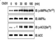
Applications : Cell assay
Review: L6 cells were incubated with CHI3L1 (100 ng/mL) for the indicated times. Cell lysates were analyzed by Western blot using antibodies against phospho-AMPKα (Thr172) and phospho-ACC (Ser79).
By Anonymous
If I order Lyophilized form.
What should they use for solvent

