Call us
301-363-4651 (Available 9 a.m. to 5 p.m. CST from Monday to Friday)
| Code | CSB-EP026136HU |
| Abbreviation | Recombinant Human WNT3A protein |
| MSDS | |
| Size | US$256 |
| Order now | |
| Image |
|
| Have Questions? | Leave a Message or Start an on-line Chat |
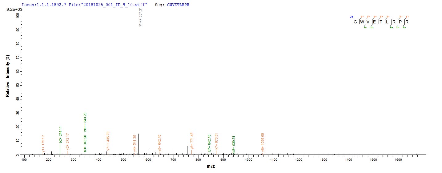
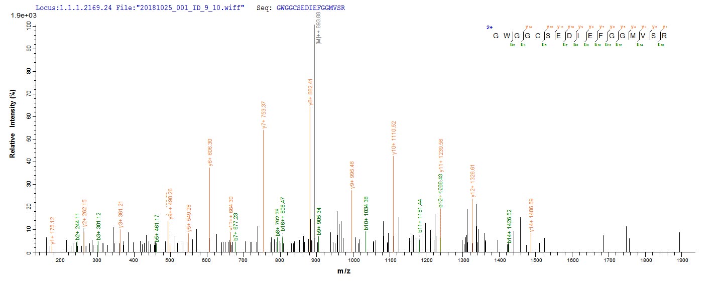
The production of recombinant human WNT3A begins with the isolation and cloning of the gene encoding the WNT3A protein (19-352aa). This gene is inserted into an expression vector along with the N-terminal 10xHis-tag gene and transfected into E.coli cells. These cells are cultured in bioreactors to express the WNT3A protein. After cell growth, the WNT3A protein is extracted and purified using the affinity chromatography technique. The purified WNT3A protein is subjected to an SDS-PAGE test, with purity of over 85%.
WNT3A is an endogenous pro-regenerative protein found in the central nervous system (CNS) [1]. WNT3A regulates RhoA-GTP levels by engaging Fzd receptors to modulate the interaction between the cytoplasmic WNT signaling protein Dishevelled (Dvl) and the formin protein Daam‐1 [2]. WNT3A stimulates the Wnt/β-catenin pathway and functions as a positive regulator of autophagy, enhancing radioresistance in squamous cell carcinoma of the head and neck [3]. It has been demonstrated that WNT3A can activate pertussis toxin (PTX)-sensitive G proteins through specific FZD receptors [4].
WNT3A is associated with dorsal-ventral patterning in the inner ear and has been shown to induce BMP-4 to specify slow myofibrogenesis of fetal myoblasts [5][6]. Research showed that WNT3A inhibited follicle-stimulating hormone-mediated steroidogenesis in primary cultures of rat granulosa cells [7]. The WNT3A signal is predominantly transduced through β‐catenin but also through the Yap/Taz pathway independently of β‐catenin, regulating various biological processes such as osteogenic differentiation, gene expression, and cell migration [8].
References:
[1] Z. Wei, J. Zhang, T. Taylor, X. Gu, Y. Zhao, & L. Wei, Neuroprotective and regenerative roles of intranasal wnt-3a administration after focal ischemic stroke in mice, Journal of Cerebral Blood Flow & Metabolism, vol. 38, no. 3, p. 404-421, 2017. https://doi.org/10.1177/0271678x17702669
[2] B. Steele, M. Harper, A. Smolenski, N. Alkazemi, A. Poole, D. Fitzgeraldet al., Wnt‐3a modulates platelet function by regulating small gtpase activity, Febs Letters, vol. 586, no. 16, p. 2267-2272, 2012. https://doi.org/10.1016/j.febslet.2012.05.060
[3] Q. Jing, G. Li, X. Chen, C. Liu, S. Lu, H. Zhenget al., Wnt3a promotes radioresistance via autophagy in squamous cell carcinoma of the head and neck, Journal of Cellular and Molecular Medicine, vol. 23, no. 7, p. 4711-4722, 2019. https://doi.org/10.1111/jcmm.14394
[4] M. Kilander, C. Halleskog, & G. Schulte, Recombinant wnts differentially activate β‐catenin‐dependent and ‐independent signalling in mouse microglia‐like cells, Acta Physiologica, vol. 203, no. 3, p. 363-372, 2011. https://doi.org/10.1111/j.1748-1716.2011.02324.x
[5] C. Forristall, F. Stellabotte, A. Castillo, & A. Collazo, Embryological manipulations in the developing xenopus inner ear reveal an intrinsic role for wnt signaling in dorsal–ventral patterning, Developmental Dynamics, vol. 243, no. 10, p. 1262-1274, 2014. https://doi.org/10.1002/dvdy.24116
[6] K. Kuroda, S. Kuang, M. Taketo, & M. Rudnicki, Canonical wnt signaling induces bmp-4 to specify slow myofibrogenesis of fetal myoblasts, Skeletal Muscle, vol. 3, no. 1, 2013. https://doi.org/10.1186/2044-5040-3-5
[7] A. Stapp, B. Gómez, C. Gifford, D. Hallford, & J. Gifford, Canonical wnt signaling inhibits follicle stimulating hormone mediated steroidogenesis in primary cultures of rat granulosa cells, Plos One, vol. 9, no. 1, p. e86432, 2014. https://doi.org/10.1371/journal.pone.0086432
[8] R. Chalamalasetty, R. Ajima, R. Garriock, M. Kennedy, L. Tessarollo, & T. Yamaguchi, A new gain‐of‐function mouse line to study the role of wnt3a in development and disease, Genesis, vol. 54, no. 9, p. 497-502, 2016. https://doi.org/10.1002/dvg.22959
There are currently no reviews for this product.

