
Call us
301-363-4651 (Available 9 a.m. to 5 p.m. CST from Monday to Friday)

| Code | CSB-RA130615A0HU |
| Size | US$210 |
| Order now | |
| Image |
|
| Have Questions? | Leave a Message or Start an on-line Chat |
| Application | Recommended Dilution |
|---|---|
| WB | 1:500-1:5000 |
| IHC | 1:50-1:200 |
| IF | 1:50-1:200 |
| FC | 1:50-1:200 |
PIK3R4, also known as VPS15 or PI3-kinase p150 subunit, serves as a critical regulatory component of the class III phosphatidylinositol 3-kinase complex. This protein plays essential roles in autophagy initiation, endosomal trafficking, and cellular homeostasis, making it a key target for researchers investigating membrane dynamics, nutrient sensing pathways, and autophagy-related disease mechanisms.
This recombinant monoclonal antibody, generated against a synthetic peptide derived from human PIK3R4, offers the reproducibility and consistency that demanding experimental workflows require. As a sequence-defined recombinant clone, it eliminates the lot-to-lot variability inherent in traditional hybridoma-derived antibodies, ensuring your results remain comparable across extended studies and collaborative projects.
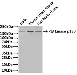
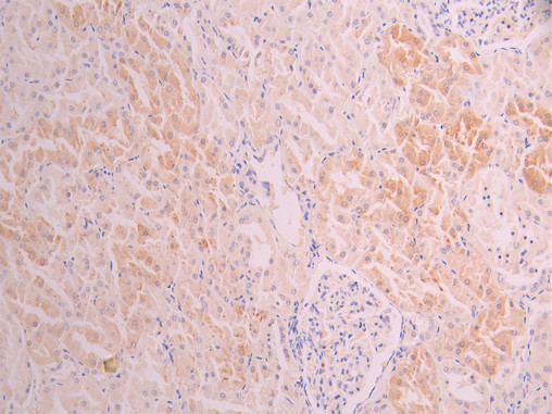
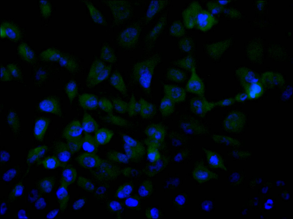
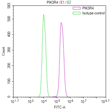
Validation data demonstrates robust performance across multiple detection platforms. Western blot analysis confirms specific detection at the expected 153 kDa molecular weight in HeLa whole cell lysates, with additional cross-species reactivity observed in mouse and rat brain tissue lysates, supporting translational research applications. Immunohistochemistry staining in paraffin-embedded human kidney tissue reveals clear target localization using standard citrate-based antigen retrieval protocols. Immunofluorescence studies in MCF-7 cells show distinct cytoplasmic staining patterns consistent with PIK3R4's known subcellular distribution. Flow cytometry validation in A431 cells demonstrates clear positive population shifts compared to isotype controls, confirming utility for single-cell analysis applications.
This versatile antibody supports researchers studying autophagy regulation, PI3K signaling networks, vesicular trafficking, and related cellular processes. Its validated performance across immunohistochemistry, immunofluorescence, and flow cytometry provides flexibility for both tissue-based and cell-based experimental approaches, while the unconjugated format allows pairing with your preferred secondary detection systems.
There are currently no reviews for this product.