The Kallikrein (KLK) Family:
Nomenclature, Cascades,
Disease Roles & Research Products

At the 2026 ASCO Annual Meeting, Johnson & Johnson announced Phase 1b data for pasritamig, a KLK2/CD3 bispecific TCE, in combination with docetaxel for metastatic castration-resistant prostate cancer (mCRPC): the combination showed significant efficacy with a favorable safety profile and no cytokine release syndrome of any grade, and has advanced to Phase 3 clinical trials. KLK2 has transitioned from a “classic prostate cancer biomarker” to a next-generation immunotherapy star target, bringing the KLK family back into the research spotlight.

The human tissue kallikrein-related peptidase (KLK) family comprises 15 secreted serine proteases and forms the largest contiguous protease gene cluster in the human genome. Beyond prostate cancer, KLK family members play important roles in various diseases, including inflammatory skin disorders, neurological diseases, and ovarian cancer. This article systematically reviews the biological characteristics, disease associations, and research progress of the KLK family to provide a reference for researchers in related fields.

1. Overview of the KLK Family

Family Members and Gene Structure

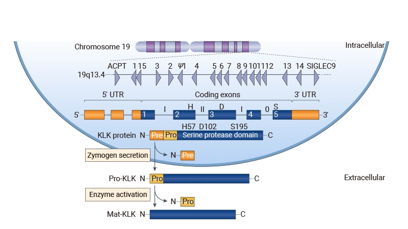
The human tissue kallikrein family consists of 15 secreted serine proteases (KLK1–KLK15), whose encoding genes are clustered on the long arm of chromosome 19 at 19q13.4, forming the largest contiguous protease gene cluster in the human genome. All KLKs are single-chain polypeptides, sharing approximately 40% sequence homology among family members, with KLK2 and KLK3 showing the highest homology (~80%) [1].

Taxonomically, KLKs belong to the S1 family of serine proteases (chymotrypsin-like), which includes approximately 80% of all known human serine proteases. In addition to tissue kallikreins, humans also have plasma kallikrein (KLKB1), encoded by the KLKB1 gene on chromosome 4, which does not belong to the KLK family [2].

Figure 1. Schematic representation of KLK family gene and protein structure

Figure 1. Schematic representation of KLK family gene and protein structure

Source: Prassas I, et al. Nat Rev Drug Discov. 2015;14(12):832-846.

Protein Structure and Zymogen Activation

All KLKs are synthesized and secreted as zymogens (pro-KLKs), comprising an N-terminal signal peptide, a propeptide, and a mature protease domain. The signal peptide is cleaved during secretion, and the propeptide is removed extracellularly via proteolytic cleavage to generate the active form.

Activation cleavage typically occurs after basic amino acids (Lys or Arg) and is mediated by trypsin-like activity. Most KLKs can be activated by other KLKs, and some (such as KLK2, KLK5, KLK6, KLK13) are capable of autoactivation. The sole exception is KLK4, whose activation requires cleavage after Gln30, mediated by a metalloproteinase [2].

Structurally, KLKs exhibit a typical serine protease fold — two β-barrel domains interact to form the active site cleft, with the catalytic triad (His57-Asp102-Ser195, chymotrypsinogen numbering) located at the bottom of the cleft. KLK1–KLK3 feature a prominent 99-loop (also called the kallikrein loop) near the substrate binding site, a characteristic structural feature of the KLK family that participates in substrate specificity regulation and endogenous inhibitor binding.

Activity Regulation Mechanisms

The proteolytic activity of KLKs is tightly regulated at multiple levels:

Regulatory Level Regulatory Mechanism Key Factors
Transcriptional Hormonal regulation, DNA methylation, miRNA Androgens, glucocorticoids, vitamin D [1] [3]
Post-translational Glycosylation N-glycosylation/O-glycosylation, affecting substrate binding and inhibitor interactions [4]
Zymogen activation Cascade activation / autoactivation KLK cascade network (tissue-specific) [5]
Activity inhibition Endogenous protein inhibitors LEKTI (skin), serpins, α2-macroglobulin, Zn²⁺ [2] [6]

Among endogenous inhibitors, LEKTI (encoded by the SPINK5 gene) is a key regulator of KLK activity in the skin, controlling the activities of KLK5, KLK7, KLK14, and others through reversible and allosteric binding. Zn²⁺ ions are also important physiological inhibitors — in the prostate, high Zn²⁺ concentrations suppress KLK activity; after ejaculation, Zn²⁺ transfers from KLK to semenogelins, releasing active KLKs and liquefying the seminal clot [2].

Tissue Distribution and Disease Associations of Family Members

Target Primary Tissue Distribution Major Disease Associations
KLK1 Kidney, pancreas, salivary glands, prostate Acute ischemic stroke, hypertension, asthma
KLK2 Prostate Prostate cancer
KLK3 Prostate Prostate cancer
KLK4 Prostate, teeth Prostate cancer, enamel formation
KLK5 Skin, breast, brain Netherton syndrome, atopic dermatitis
KLK6 Brain, ovary, breast Multiple sclerosis, Alzheimer’s disease, ovarian cancer
KLK7 Skin, esophagus, kidney Netherton syndrome, atopic dermatitis, colon cancer
KLK8 Skin, brain, ovary Psoriasis, neural plasticity, ovarian cancer
KLK9 Thymus, testis Breast cancer, ovarian cancer
KLK10 Breast, prostate, ovary Breast cancer, ovarian cancer
KLK11 Prostate, ovary, stomach Ovarian cancer, gastric cancer
KLK12 Salivary glands, stomach Less studied, angiogenesis
KLK13 Thyroid, breast, prostate Thyroid cancer, liver cancer
KLK14 Skin, prostate, breast Netherton syndrome, prostate cancer, renal cancer
KLK15 Prostate, thyroid Prostate cancer, renal cancer

2. Prostate Cancer

Prostate cancer is the most extensively studied disease area for the KLK family. From the decades-long clinical application of KLK3 (PSA) as the “gold standard” biomarker to the recent transformation of KLK2 from biomarker to therapeutic target, the KLK family has consistently been one of the core target groups in prostate cancer research.

KLK2

KLK2 (human glandular kallikrein) shares approximately 80% amino acid homology with KLK3 (PSA), and their genes are located only ~12 kb apart in the 19q13.4 region [1]. Traditionally, KLK2 was considered a secreted protease whose primary function is to activate pro-KLK3 (PSA) and participate in semen liquefaction.

In 2025, Shen et al. published a study in Clinical Cancer Research that, for the first time, demonstrated KLK2 expression on the surface of prostate cancer cells: using FACS and confocal microscopy, the researchers detected KLK2 signals on the cell membrane in prostate cancer cell lines and patient-derived tumors [7]. This finding changed the traditional view of KLK as a purely secreted protease and provided key structural biology support for the development of antibody-based drugs targeting KLK2.

Drug Pipeline Progress:

Drug Name Target Indication Modality Stage Company
Pasritamig KLK2×CD3 Prostate cancer Bispecific TCE Phase 3 Johnson & Johnson
Ac-225-labeled anti-KLK2 antibody KLK2 Prostate cancer Radioimmunotherapy Phase 1 Johnson & Johnson
JNJ-75229414 KLK2 Prostate cancer CAR-T Phase 1 Johnson & Johnson
JNJ-98768111 KLK2 Prostate cancer ADC Phase 1 Johnson & Johnson

Pasritamig is a first-in-class KLK2-targeted bispecific antibody whose differentiated advantage lies in the high tissue specificity of KLK2 — KLK2 is almost exclusively expressed in prostate tissue, with very low expression in other tissues, which significantly reduces the risk of off-target toxicity compared to PSMA-targeted agents (PSMA is expressed in salivary glands, kidneys, and nervous tissue, often causing adverse effects such as dry mouth) [11].

KLK3/PSA

KLK3 encodes prostate-specific antigen (PSA), the “gold standard” clinical biomarker for prostate cancer screening, with chymotrypsin-like specificity. The classic function of PSA is to hydrolyze semenogelins I/II and fibronectin, leading to liquefaction of the seminal clot and release of sperm [2].

For a long time, KLK3/PSA was primarily used as a diagnostic biomarker. In recent years, with deeper understanding of its biological functions, KLK3 has been gradually transitioning from biomarker to therapeutic target:

Drug Pipeline Progress:

Drug Name Target Indication Modality Stage Company
Afala KLK3 Prostate cancer Monoclonal antibody Approved (2014) Materia Medica Holding
RAD 402 KLK3 Prostate cancer Antibody-conjugated radionuclide Phase 1/2 Radiopharm Theranostics
HB-300 ACP3×KLK3 Prostate cancer Recombinant vector vaccine Phase 1/2 HOOKIPA Pharma

RAD 402 (NCT07259213) dosed its first patient in March 2026 and represents a representative advance in KLK3-targeted radioligand therapy [12]. Therapeutic cancer vaccines (such as HB-300) use viral vectors to express PSA antigen in vivo, eliciting CTL responses to kill PSA-positive prostate cancer cells [13].

Since KLK2 and KLK3 share 80% amino acid homology and PSA circulates at high concentrations in the blood, the risk of cross-reactivity is an unavoidable concern when developing KLK2-targeted antibodies. Studies have shown that the high homology between KLK2 and KLK3 leads to a significantly higher probability of antibody cross-binding compared to other family members, so cross-reactivity testing against KLK3 requires special attention during antibody screening and specificity evaluation [7].

KLK4

Unlike other KLKs (which are all secreted extracellular proteins), KLK4 is primarily localized to the nucleus. The full-length hK4-254 is cytoplasmic, while the N-terminally truncated hK4-205 is nuclear. Nuclear-localized KLK4 promotes prostate cancer cell proliferation by regulating transcription factors such as p21 and cyclin D1 [14].

This property means KLK4 is not suitable as a cell surface target for antibody drugs; currently, only early-stage explorations exist with cell therapies such as TCR-T [15].

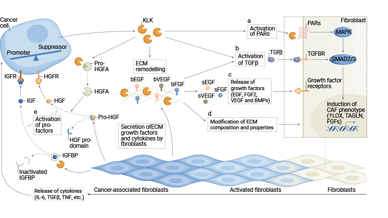
Figure 2. Multiple mechanisms by which the KLK family remodels the tumor microenvironment. KLKs regulate tumor cell–microenvironment interactions through degrading ECM proteins, cleaving cell adhesion molecules, releasing ECM-bound growth factors, and activating other proteases.

Figure 2. Multiple mechanisms by which the KLK family remodels the tumor microenvironment. KLKs regulate tumor cell–microenvironment interactions through degrading ECM proteins, cleaving cell adhesion molecules, releasing ECM-bound growth factors, and activating other proteases.

Source: Srinivasan S, et al. Nat Rev Cancer. 2022.

3. Inflammatory Skin Diseases

The stratum corneum of the skin is another hot area of KLK family research. In the epidermis, KLK5, KLK7, and KLK14 form a precisely regulated proteolytic cascade network responsible for normal stratum corneum desquamation. When this cascade becomes dysregulated, it leads to a range of inflammatory skin diseases.

The Skin KLK Proteolytic Cascade

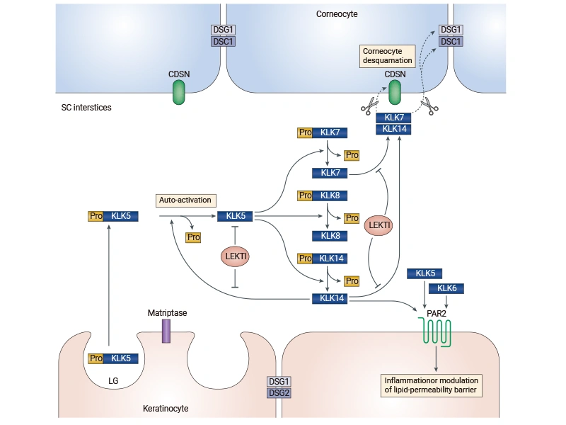
In normal skin, during differentiation, keratinocytes secrete inactive pro-KLKs into the intercellular spaces of the stratum corneum via lamellar bodies (LGs). These zymogens form an activation cascade:

Figure 3. Schematic representation of the skin epidermal KLK proteolytic cascade

Figure 3. Schematic representation of the skin epidermal KLK proteolytic cascade

Source: Prassas I, et al. Nat Rev Drug Discov. 2015.

KLK5 is the initiator of the cascade — proKLK5 possesses autoactivation capacity and can convert itself to the active form, subsequently cleaving and activating proKLK7, proKLK8, and proKLK14. Active KLK14 can in turn activate proKLK5, creating a positive feedback loop that amplifies the proteolytic cascade. Activated KLK5, KLK7, and KLK14 ultimately degrade corneodesmosomal proteins (DSG1, DSC1, CDSN), leading to breakdown of intercellular connections between corneocytes and normal desquamation [16].

The activity of this cascade is finely regulated by multiple factors. Within the epidermis, there exist pH and calcium gradients: the calcium gradient controls the formation and secretion of lamellar bodies — through which KLK zymogens are released into the stratum corneum [36]; while the pH gradient regulates activity levels by influencing the binding of KLKs to endogenous inhibitors such as LEKTI [17]. The LEKTI protein, encoded by the SPINK5 gene, is the most critical endogenous inhibitor of KLK activity in the skin, regulating the activity of the entire cascade through reversible and allosteric binding [6].

Netherton Syndrome

Netherton syndrome (NS) is a rare inherited skin disease caused by loss-of-function mutations in the SPINK5 gene, leading to defective or absent LEKTI protein. Loss of LEKTI results in uncontrolled activation of KLK5 and downstream KLKs, causing excessive degradation of desmosomal proteins and severe impairment of the epidermal barrier [18].

NS patients present with congenital ichthyosiform erythroderma, bamboo hair (trichorrhexis invaginata), and atopic diathesis (allergic rhinitis, asthma, food allergies, etc.). Severe cases can be life-threatening due to infections and electrolyte disturbances. The incidence of NS is approximately 1 in 200,000 newborns.

Gene knockout studies have shown that in Spink5-deficient mice, simultaneous knockout of KLK5 and KLK7 can fully rescue the lethal phenotype [19]. This indicates that KLK5 is the core driver in the pathogenesis of NS, and inhibiting KLK5 is a key therapeutic strategy for NS.

Atopic Dermatitis

In lesions of patients with atopic dermatitis (AD), multiple KLKs (such as KLK5, KLK7, KLK14, etc.) are upregulated [20]. Increased KLK activity leads to abnormal epidermal barrier function, while simultaneously promoting inflammation through activation of protease-activated receptor 2 (PAR2), inducing the release of pro-inflammatory cytokines such as thymic stromal lymphopoietin (TSLP) [21].

AD is a chronic inflammatory skin disease affecting approximately 20% of children and 10% of adults globally, with a huge market size (dupilumab sales exceeded $10 billion in 2023). KLK5/7-targeted drugs offer a differentiated therapeutic mechanism for AD.

Drug Pipeline Progress:

Drug Name Target Indication Modality Stage Company
TRIV-509 KLK5×KLK7 Atopic dermatitis, asthma Bispecific antibody Phase 2 Triveni Bio
SXR-1096 KLK5×KLK7×KLK14 Netherton syndrome Small-molecule inhibitor Phase 1/2 Sixera Pharma
BCX-17725 KLK5 Netherton syndrome Fusion protein Phase 1 BioCryst
DS-2325 KLK5 Netherton syndrome Gene therapy Phase 1 Daiichi Sankyo
ATR-12 KLK5 Netherton syndrome Engineered bacteria Phase 1 Azitra

TRIV-509 is the most advanced bispecific antibody in the KLK field. It has shown superior efficacy compared to IL-4R inhibitors (dupilumab) in multiple preclinical AD models and demonstrated rapid improvement in skin biopsy samples from AD patients [22]. Phase II clinical trials are currently underway.

4. Neuroscience and Stroke

The role of the KLK family in the nervous system is equally important. KLK1, KLK6, and KLK8 are the three most extensively studied members in the brain, involved in stroke neuroprotection, demyelinating diseases, and neural plasticity, respectively.

KLK1

KLK1 (tissue kallikrein) is the earliest discovered and most extensively studied member of the KLK family. Its classic function is to catalyze the release of kinins (bradykinin and kallidin) from kininogens, participating in blood pressure regulation, inflammatory responses, and tissue repair through the kinin B2 receptor signaling pathway [23].

In acute ischemic stroke (AIS), KLK1 has well-documented neuroprotective effects, improving cerebral blood flow, reducing infarct volume, and promoting neurological recovery [24].

Drug Pipeline Progress:

Drug Name Target Indication Modality Stage Company
DM199 KLK1 Acute ischemic stroke Recombinant protein Phase 2/3 DiaMedica
PTH-0366 KLK1 Acute ischemic stroke Recombinant protein Phase 1 Protheragen

DM199 (recombinant human tissue kallikrein 1) has entered a global multicenter Phase II/III clinical trial (NCT05065216), enrolling patients at over 100 research centers worldwide [25].

KLK6

KLK6 (also known as Neurosin, Zyme) is one of the most abundant KLKs in the brain and spinal cord. It is synthesized de novo by reactive astrocytes and is a molecular trigger of reactive astrogliosis.

KLK6 is closely associated with inflammatory demyelination in multiple sclerosis (MS) lesions. Inhibition of KLK6 delays symptom onset in experimental autoimmune encephalomyelitis (EAE). In Alzheimer’s disease, KLK6 participates in amyloid metabolism and may affect the production and clearance of Aβ [26].

Currently, KLK6-targeted drugs are still in the preclinical stage, including engineered protein inhibitors, small-molecule inhibitors, and covalent probes [27].

KLK8

KLK8 (also known as Neuropsin) is a key regulator of neural plasticity. It is highly expressed in the hippocampus and participates in synaptic remodeling during learning and memory. KLK8 cleaves extracellular matrix proteins and neural cell adhesion molecules to regulate synaptic plasticity [28].

In the skin, KLK8 is significantly upregulated in psoriatic lesions, and serum KLK8 levels correlate with disease severity. KLK8 participates in the formation of psoriatic lesions, playing a particularly key role in the development of neutrophil microabscesses, but its mechanism of action differs from that of classic desquamatory KLKs [29].

5. Cancer Biomarkers

Beyond the three main research areas outlined above, multiple KLK family members hold significant value as biomarkers in various cancers:

Ovarian cancer:

KLK6, KLK10, KLK11, and others are aberrantly expressed in ovarian cancer and serve as diagnostic and prognostic biomarkers. KLK6 is elevated in the ascites of patients with peritoneal metastasis and can be used to differentiate benign from malignant ascites [30]. KLK11 can distinguish ovarian cancer from healthy controls and is suitable for longitudinal screening [31].

Breast cancer:

KLK10 acts as a tumor suppressor in breast cancer, where promoter methylation leads to silencing of expression, associated with poor prognosis [32]. KLK6 promotes breast cancer cell invasion and is associated with metastasis [30].

Colon cancer:

KLK6 and KLK7 are significantly upregulated in colon cancer. KLK7 promotes invasion and peritoneal dissemination of colorectal cancer cells and exhibits high diagnostic efficacy [33]. KLK10 plays an oncogenic role in colon cancer with significantly elevated expression [30].

Other cancers:

KLK13 is aberrantly expressed in thyroid cancer and has potential as a diagnostic biomarker [30]. KLK15 is upregulated in prostate cancer, associated with tumor aggressiveness, and can serve as a prognostic indicator [35].

CUSABIO KLK Family Products

To support KLK family-related research, CUSABIO provides the following recombinant proteins, ELISA kits, and antibody products:

● Recombinant Protein

Target Code Product Name Source
KLK1 CSB-EP012446HU Recombinant Human Kallikrein-1 (KLK1) E.coli
KLK10 CSB-EP012447HU1 Recombinant Human Kallikrein-10 (KLK10), partial E.coli
KLK2 CSB-EP012453HU Recombinant Human Kallikrein-2 (KLK2) E.coli
KLK7 CSB-EP012458HU Recombinant Human Kallikrein-7 (KLK7), partial E.coli
Klk7 CSB-EP012458RA Recombinant Rat Glandular kallikrein-7, submandibular/renal (Klk7) E.coli
Klk1b22 CSB-EP325391MO Recombinant Mouse Kallikrein 1-related peptidase b22 (Klk1b22) E.coli
Klk8 CSB-EP720268MO Recombinant Mouse Kallikrein-8 (Klk8) E.coli
Klk14 CSB-EP806542MO Recombinant Mouse Kallikrein-14 (Klk14) E.coli
Klk14 CSB-EP806542MOa2 Recombinant Mouse Kallikrein-14 (Klk14) E.coli
KLK7 CSB-YP012458HU Recombinant Human Kallikrein-7 (KLK7), partial Yeast
Klk8 CSB-YP720268MO Recombinant Mouse Kallikrein-8 (Klk8) Yeast
Klk14 CSB-YP806542MO Recombinant Mouse Kallikrein-14 (Klk14) Yeast
KLK9 CSB-MP892334HU Recombinant Human Kallikrein-9 (KLK9) Mammalian cell
KLK15 CSB-MP752074SWB Recombinant Saguinus oedipus Kallikrein-15 (KLK15) Mammalian cell
KLK9 CSB-EP892334HU Recombinant Human Kallikrein-9 (KLK9) E.coli
KLK12 CSB-EP892137HU Recombinant Human Kallikrein-12 (KLK12) E.coli
KLK7 CSB-EP012458HU1 Recombinant Human Kallikrein-7 (KLK7), partial E.coli
Klk1b5 CSB-EP325792MO Recombinant Mouse Kallikrein 1-related peptidase b5 (Klk1b5) E.coli
KLK3 CSB-EP012454MOW Recombinant Macaca mulatta Prostate-specific antigen (KLK3) E.coli
KLK2 CSB-YP012453HU Recombinant Human Kallikrein-2 (KLK2) Yeast

● Antibody

Target Code Product Name Tested Applications
KLK3 CSB-RA548583A0HU KLK3 Recombinant Monoclonal Antibody ELISA, IHC; Recommended dilution: IHC:1:50-1:200
KLK5 CSB-RA729707A0HU KLK5 Recombinant Monoclonal Antibody ELISA, WB, IHC, FC; Recommended dilution: WB:1:500-1:5000, IHC:1:50-1:200, FC:1:20-1:200
KLK2 CSB-RA012453MA1HU KLK2 Recombinant Monoclonal Antibody ELISA; Recommended dilution: ELISA:1:1000-1:10000
KLK11 CSB-PA000045 Cleaved-KLK11 (I54) Antibody WB, ELISA; WB:1:500-1:2000, ELISA:1:10000
KLK8 CSB-PA000046 Cleaved-KLK8 (V33) Antibody WB, ELISA; WB:1:500-1:2000, ELISA:1:10000
KLK1 CSB-PA005798 KLK1 Antibody WB, ELISA; WB:1:500-1:2000, ELISA:1:20000
KLK3 CSB-PA006113 KLK3 Antibody WB, ELISA; WB:1:500-1:2000, ELISA:1:10000
KLK3 CSB-PA009525 KLK3 Antibody WB, IHC, IF, ELISA; WB:1:500-1:2000, IHC:1:100-1:300, IF:1:200-1:1000, ELISA:1:20000
KLK1 CSB-PA012446GA01HU KLK1 Antibody ELISA, WB, IHC
KLK11 CSB-PA012448GA01HU KLK11 Antibody ELISA, IHC
KLK5 CSB-PA012456GA01HU KLK5 Antibody ELISA
KLK8 CSB-PA012459GA01HU KLK8 Antibody ELISA, WB
KLK3 CSB-PA012454GA01HU KLK3 Antibody ELISA, IHC
KLK3 CSB-PA920807 KLK3 Antibody ELISA, IHC; ELISA:1:500-1:2000, IHC:1:5-1:20
KLK3 CSB-PA554992 KLK3 Antibody ELISA, IHC; ELISA:1:500-1:2000, IHC:1:10-1:25
KLK1 CSB-PA086611 KLK1 Antibody ELISA, IHC; ELISA:1:1000-1:5000, IHC:1:50-1:200
KLK1 CSB-PA248038 KLK1 Antibody ELISA, IHC; ELISA:1:1000-1:5000, IHC:1:50-1:200
KLK4 CSB-PA211882 KLK4 Antibody ELISA, IHC; ELISA:1:2000-1:5000, IHC:1:25-1:100
KLK4 CSB-PA214768 KLK4 Antibody ELISA, IHC; ELISA:1:2000-1:5000, IHC:1:25-1:100
KLK7 CSB-PA094965 KLK7 Antibody ELISA, IHC; ELISA:1:1000-1:5000, IHC:1:25-1:100

● ELISA Kit

Target Code Product Name Detection Range Sensitivity
KLK3 CSB-E08276m Mouse Kallikrein 1-related peptidase b3,Klk3 ELISA Kit 0.312 ng/mL-20 ng/mL 0.078 ng/mL
KLK6 CSB-E09248h Human Kallikrein 6,KLK 6 ELISA Kit 1.25 ng/ml-80 ng/ml 0.31 ng/mL
KLK10 CSB-E10095h Human Kallikrein 10,KLK 10 ELISA Kit 1.675 ng/mL-40 ng/mL 0.7 ng/mL
KLK1 CSB-E11437h Human Kallikrein 1,KLK 1 ELISA Kit 0.78 ng/mL-50 ng/mL 0.195 ng/mL
KLK2 CSB-E13094h Human Kallikrein 2(hK 2) ELISA Kit 62.5 pg/mL-4000 pg/mL 15.6 pg/mL
KLK7 CSB-E13797h Human Kallikrein 7,KLK 7 ELISA Kit 0.625 ng/mL-40 ng/mL 0.156 ng/mL
KLK14 CSB-EL012451HU Human Kallikrein-14(KLK14) ELISA kit 1.56 ng/ml-100 ng/ml 0.39 ng/ml
KLK4 CSB-EL012455HU Human Kallikrein-4(KLK4) ELISA kit 0.156 ng/mL-10 ng/mL 0.039 ng/mL
KLK5 CSB-EL012456HU Human Kallikrein-5(KLK5) ELISA kit 62.5 pg/mL-4000 pg/mL 15.6 pg/mL

References

[1] Yousef GM, Diamandis EP. The new human tissue kallikrein gene family: structure, function, and association to disease. Endocr Rev. 2001;22 (2):184-204.

[2] Prassas I, Eissa A, Poda G, et al. Unleashing the therapeutic potential of human kallikrein-related serine proteases. Nat Rev Drug Discov. 2015;14 (3):183-202.

[3] Shaw JL, Diamandis EP. Distribution of 15 human kallikreins in tissues and biological fluids. Clin Chem. 2007;53 (8):1423-32.

[4] Kalinska M,Meyer-Hoffert U,Kantyka T, et al. Kallikreins - The melting pot of activity and function. Biochimie. 2016;122:270-82.

[5] Pampalakis G, Sotiropoulou G. Tissue kallikrein proteolytic cascade pathways in normal physiology and cancer. Biochim Biophys Acta. 2007;1776 (1):22-31.

[6] Hovnanian A. Netherton syndrome: skin inflammation and allergy by loss of protease inhibition. Cell Tissue Res. 2013;351 (2):289-300.

[7] Shen F, Smith R, McDevitt T, et al. Human Kallikrein 2: A Novel Lineage-Specific Surface Target in Prostate Cancer. Clin Cancer Res. 2025;31 (21):4543-4556.

[8] URL: https://www.jnj.com/media-center/press-releases/johnson-johnson-ushers-in-the-next-wave-of-innovation-in-cancer-care-with-more-than-20-clinical-and-real-world-studies-at-asco-2026

[9] URL: https://www.asco.org/abstracts-presentations/256425

[10] Blinka S, Yu EY. Drug Targets in Prostate Cancer: An Appetite for KLK2-Mediated Destruction. Clin Cancer Res. 2025;31 (21):4393-4395.

[11] Subudhi SK, Siddiqui BA, Zarrabi KK, et al. The path forward for T cell engagers in patients with prostate cancer. J Immunother Cancer. 2026;14 (2):.

[12] URL: https://clinicaltrials.gov/study/NCT07259213

[13] HB-302/​HB-301 Therapy in Participants With Metastatic Castration-Resistant Prostate Cancer. ClinicalTrials.gov. 2026. URL: https://clinicaltrials.gov/study/NCT05553639

[14] Klokk TI, Kilander A, Xi Z, et al. Kallikrein 4 is a proliferative factor that is overexpressed in prostate cancer. Cancer Res. 2007;67 (11):5221-30.

[15] van Amerongen RA, Tuit S, Remst DFG, et al. TCR-Based Therapy Directed against Kallikrein-Related Peptidase 4 Is Safe and Effective against Prostate Cancer. Cancer Immunol Res. 2025;13 (8):1145-1159.

[16] Brattsand M,Stefansson K,Lundh C, et al. A proteolytic cascade of kallikreins in the stratum corneum. J Invest Dermatol. 2005;124 (1):198-203.

[17] Nauroy P, Nyström A. Kallikreins: Essential epidermal messengers for regulation of the skin microenvironment during homeostasis, repair and disease. Matrix Biol Plus. 2020;6-7:100019.

[18] Chavanas S,Bodemer C,Rochat A, et al. Mutations in SPINK5, encoding a serine protease inhibitor, cause Netherton syndrome. Nat Genet. 2000;25 (2):141-2.

[19] Kasparek P,Ileninova Z,Zbodakova O, et al. KLK5 and KLK7 Ablation Fully Rescues Lethality of Netherton Syndrome-Like Phenotype. PLoS Genet. 2017;13 (1):e1006566.

[20] Zani MB, Sant'Ana AM, Tognato RC, et al. Human Tissue Kallikreins-Related Peptidases Are Targets for the Treatment of Skin Desquamation Diseases. Front Med (Lausanne). 2021;8:777619.

[21] Briot A, Deraison C, Lacroix M, et al. Kallikrein 5 induces atopic dermatitis-like lesions through PAR2-mediated thymic stromal lymphopoietin expression in Netherton syndrome. J Exp Med. 2009;206 (5):1135-47.

[22] URL: https://triveni.bio/pipeline/

[23] Clements JA,Willemsen NM,Myers SA, et al. The tissue kallikrein family of serine proteases: functional roles in human disease and potential as clinical biomarkers. Crit Rev Clin Lab Sci. 2004;41 (3):265-312.

[24] URL: https://www.diamedica.com/our-focus

[25] URL: https://clinicaltrials.gov/study/NCT05065216

[26] Mella C,Figueroa CD,Otth C, et al. Involvement of Kallikrein-Related Peptidases in Nervous System Disorders. Front Cell Neurosci. 2020;14:166.

[27] Dreyer T,Dreyer T,Dreyer T, et al. Drug Discovery Strategies for Kallikrein-Related Peptidases. Int J Mol Sci. 2025;27 (1):.

[28] Bukowski L,Chernomorchenko AMF,Starnawska A, et al. Neuropsin in mental health. J Physiol Sci. 2020;70 (1):26.

[29] Iinuma S,Kishibe M,Saito N, et al. Klk8 is required for microabscess formation in a mouse imiquimod model of psoriasis. Exp Dermatol. 2015;24 (11):887-9.

[30] Borgoño CA,Diamandis EP. The emerging roles of human tissue kallikreins in cancer. Nat Rev Cancer. 2004;4 (11):876-90.

[31] McIntosh MW,Liu Y,Drescher C, et al. Validation and characterization of human kallikrein 11 as a serum marker for diagnosis of ovarian carcinoma. Clin Cancer Res. 2007;13 (15 Pt 1):4422-8.

[32] Sidiropoulos M,Pampalakis G,Sotiropoulou G, et al. Downregulation of human kallikrein 10 (KLK10/NES1) by CpG island hypermethylation in breast, ovarian and prostate cancers. Tumour Biol. 2005;26 (6):324-36.

[33] Talieri M,Mathioudaki K,Prezas P, et al. Clinical significance of kallikrein-related peptidase 7 (KLK7) in colorectal cancer. Thromb Haemost.

[34] Obiezu CV,Diamandis EP. Human tissue kallikrein gene family: applications in cancer. Cancer Lett. 2005;224 (1):1-22.

[35] Rabien A,Fritzsche FR,Jung M, et al. KLK15 is a prognostic marker for progression-free survival in patients with radical prostatectomy. Int J Cancer. 2010;127 (10):2386-94.

[36] Leprince C,Simon M. Epidermal lamellar bodies, essential organelles for the skin barrier. Front Cell Dev Biol. 2025;13:1597884.

icon of phone
Call us
301-363-4651 (Available 9 a.m. to 5 p.m. CST from Monday to Friday)
icon of address
Address
No. 269, Shendun 5th Road, Donghu Hi-Tech Development Area, Hubei Province, 430206, P.R.China
icon of social media
Join us with

Subscribe newsletter

Leave a message

* To protect against spam, please pass the CAPTCHA test below.
CAPTCHA verification
© 2007-2026 CUSABIO TECHNOLOGY LLC All rights reserved. 鄂ICP备15011166号-1