PRKAR2A Recombinant Monoclonal Antibody

Code CSB-RA197122A0HU
Size US$210
Order now
Image
  • IHC image of CSB-RA197122A0HU diluted at 1:50 and staining in paraffin-embedded human kidney tissue performed on a Leica BondTM system. After dewaxing and hydration, antigen retrieval was mediated by high pressure in a citrate buffer (pH 6.0). Section was blocked with 10% normal goat serum 30min at RT. Then primary antibody (1% BSA) was incubated at 4°C overnight. The primary is detected by a Goat anti-rabbit polymer IgG labeled by HRP and visualized using 0.47% DAB.
  • IHC image of CSB-RA197122A0HU diluted at 1:50 and staining in paraffin-embedded human rectal cancer performed on a Leica BondTM system. After dewaxing and hydration, antigen retrieval was mediated by high pressure in a citrate buffer (pH 6.0). Section was blocked with 10% normal goat serum 30min at RT. Then primary antibody (1% BSA) was incubated at 4°C overnight. The primary is detected by a Goat anti-rabbit polymer IgG labeled by HRP and visualized using 0.47% DAB.
  • Immunofluorescence staining of Hela with CSB-RA197122A0HU at 1:5, counter-stained with DAPI. The cells were fixed in 4% formaldehyde and blocked in 10% normal Goat Serum. The cells were then incubated with the antibody overnight at 4°C. The secondary antibody was Alexa Fluor 512-congugated AffiniPure Goat Anti-Rabbit IgG(H+L).
  • Overlay Peak curve showing MCF-7 cells stained with CSB-RA197122A0HU (red line) at 1:50. The cells were fixed in 4% formaldehyde and permeated by 0.2% TritonX-100. Then 10% normal goat serum to block non-specific protein-protein interactions followed by the antibody (1µg/1*106cells) for 45min at 4℃. The secondary antibody used was FITC-conjugated Goat Anti-rabbit IgG(H+L) at 1:200 dilution for 35min at 4℃.Control antibody (green line) was rabbit IgG (1µg/1*106cells) used under the same conditions. Acquisition of >10,000 events was performed.
Have Questions? Leave a Message or Start an on-line Chat

Product Details

Uniprot No.
Target Names
Alternative Names
cAMP-dependent protein kinase type II-alpha regulatory subunit, PRKAR2A, PKR2 PRKAR2
Species Reactivity
Human
Immunogen
A synthesized peptide derived from Human PRKAR2A
Immunogen Species
Homo sapiens (Human)
Conjugate
Non-conjugated
Clonality
Monoclonal
Isotype
Rabbit IgG
Clone No.
15G8
Purification Method
Affinity-chromatography
Concentration
It differs from different batches. Please contact us to confirm it.
Buffer
Rabbit IgG in 10mM phosphate buffered saline , pH 7.4, 150mM sodium chloride, 0.05% BSA, 0.02% sodium azide and 50% glycerol.
Form
Liquid
Tested Applications
ELISA, IHC, IF, FC
Recommended Dilution
Application Recommended Dilution
IHC 1:50-1:200
IF 1:50-1:200
FC 1:50-1:200
Troubleshooting and FAQs
Storage
Upon receipt, store at -20°C or -80°C. Avoid repeated freeze.
Lead Time
Basically, we can dispatch the products out in 1-3 working days after receiving your orders. Delivery time maybe differs from different purchasing way or location, please kindly consult your local distributors for specific delivery time.
Description

PRKAR2A serves as the type II-alpha regulatory subunit of cAMP-dependent protein kinase, playing a central role in the cAMP signaling cascade that governs diverse cellular processes including metabolism, gene expression, and cell proliferation. As a key modulator of protein kinase A activity, PRKAR2A has drawn significant research interest for its involvement in cancer biology and metabolic regulation, where dysregulated cAMP signaling can drive pathological outcomes.

This recombinant monoclonal antibody, clone 15G8, offers the reproducibility and consistency that demanding research applications require. Generated against a synthetic peptide derived from human PRKAR2A and produced using recombinant technology, this antibody provides sequence-defined specificity with minimal lot-to-lot variation, ensuring your experimental results remain comparable across studies and over time.

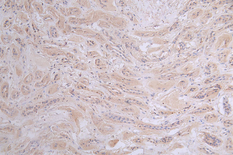
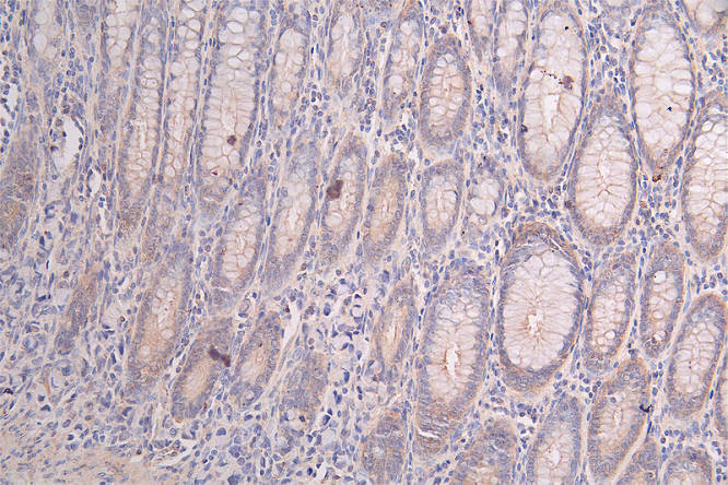
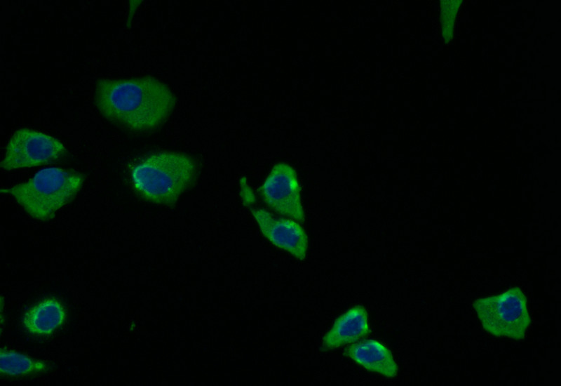
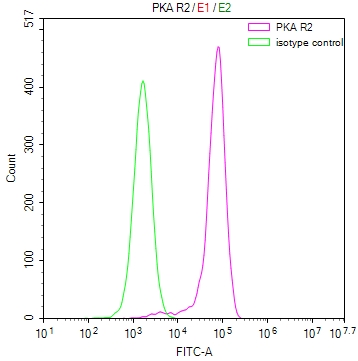
Validation across multiple detection platforms demonstrates this antibody's versatility in your workflow. Immunohistochemistry studies confirm reliable staining in paraffin-embedded human tissues, with clear detection observed in both normal kidney tissue and rectal cancer specimens at 1:50 dilution using citrate buffer antigen retrieval. For cellular localization studies, immunofluorescence analysis in HeLa cells reveals distinct staining patterns when counterstained with DAPI. Flow cytometry validation using MCF-7 breast cancer cells shows a clear positive shift compared to isotype control, confirming utility for quantitative single-cell analysis of PRKAR2A expression.

Supplied in a glycerol-containing buffer optimized for long-term stability, this antibody supports researchers investigating cAMP-dependent signaling networks, metabolic regulation, and oncogenic pathways where protein kinase A activity influences disease progression and therapeutic response.

Usage
For Research Use Only. Not for use in diagnostic or therapeutic procedures.

Customer Reviews and Q&A

 Customer Reviews

There are currently no reviews for this product.

Submit a Review here

Target Background

Function
Regulatory subunit of the cAMP-dependent protein kinases involved in cAMP signaling in cells. Type II regulatory chains mediate membrane association by binding to anchoring proteins, including the MAP2 kinase.
Gene References into Functions
  1. High PK-R2 expression is associated with colorectal cancer. PMID: 26372733
  2. Prkar2a deficiency predisposes to hematopoietic malignancies in vivo. RIIalpha's likely association with HS and DLBCL was hitherto unrecognized and may lead to better understanding of these rare neoplasms. PMID: 26608815
  3. Disruption of Snapin-PKR2 interaction did not affect PKR2 signaling, but increased the ligand-induced degradation, implying a role of Snapin in the trafficking of PKR2. PMID: 26687946
  4. we demonstrate that neurochondrin has strong isoform selectivity towards the RIIa subunit of PKA with nanomolar affinity PMID: 25916936
  5. These data demonstrate that some Kallmann syndrome-associated, intracellularly retained mutant PKR2 receptors can be functionally rescued, suggesting a potential treatment strategy for patients bearing such mutations. PMID: 24753254
  6. Smad4 and the R subunit of the protein kinase A holoenzyme form a functional complex in vivo in response to TGFbeta. PMID: 23362281
  7. ETO nervy homology region (NHR) 3 domain-PKA(RIIalpha) protein interaction does not appear to significantly contribute to AML1-ETO's ability to induce leukemia. PMID: 20708017
  8. findings indicate that increased particulate type II protein kinase A activity occurs throughout pregnancy therefore directing the cAMP quiescence signal to specific subcellular loci within myometrial smooth muscle cells PMID: 12727975
  9. These data implicate the involvement of PKA-RIIalpha anchoring apical targeting of distinct proteins and glycosphingolipids to apical plasma membrane domains and suggest that rerouting may underlie the delayed Golgi-to-apical surface transport of MDR1. PMID: 16723498
  10. The high-resolution crystal structures of the docking and dimerization (D/D) domain of the RIIalpha regulatory subunit of PKA in complex with the high-affinity anchoring peptide AKAP-IS explain the molecular basis for AKAP-regulatory subunit recognition. PMID: 17081989
  11. The data suggest that centrosomal anchoring of RIIalpha and the interrelated subapical positioning of these centrosomes is required for oncostatin M-, but not cAMP-mediated, bile canalicular lumen development. PMID: 17494870
  12. RIIalpha releases Calpha upon elevated cAMP alone, dependent on autophosphorylation of the RIIalpha inhibitory domain PMID: 17884635
  13. Bacillus anthracis edema toxin altered the protein levels and activity of protein kinase A and exchange protein activated by cAMP (Epac), a recently identified cAMP-binding molecule. PMID: 19307216

Show More

Hide All

Subcellular Location
Cytoplasm. Cell membrane. Note=Colocalizes with PJA2 in the cytoplasm and the cell membrane.
Protein Families
CAMP-dependent kinase regulatory chain family
Tissue Specificity
Four types of regulatory chains are found: I-alpha, I-beta, II-alpha, and II-beta. Their expression varies among tissues and is in some cases constitutive and in others inducible.
Database Links

HGNC: 9391

OMIM: 176910

KEGG: hsa:5576

STRING: 9606.ENSP00000265563

UniGene: Hs.631923

icon of phone
Call us
301-363-4651 (Available 9 a.m. to 5 p.m. CST from Monday to Friday)
icon of address
Address
7505 Fannin St., Ste 610, Room 7 (CUBIO Innovation Center), Houston, TX 77054, USA
icon of social media
Join us with

Subscribe newsletter

Leave a message

* To protect against spam, please pass the CAPTCHA test below.
CAPTCHA verification
© 2007-2026 CUSABIO TECHNOLOGY LLC All rights reserved. 鄂ICP备15011166号-1
Place an order now

I. Product details

*
*
*
*

II. Contact details

*
*

III. Ship To

*
*
*
*
*
*
*

IV. Bill To

*
*
*
*
*
*
*
*