-
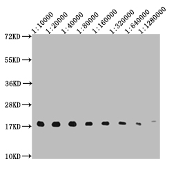
Western Blot
Positive WB detected in: 10ug Hela whole cell lysate, HistoneH3 antibody at 1:10000, 1:20000, 1:40000, 1:80000, 1:160000, 1:320000, 1:640000, 1:1280000
Secondary
Goat polyclonal to mouse IgG at 1/50000 dilution
Predicted band size: 15-25 KDa
Observed band size: 15-25 KDa
Exposure time:5s
-
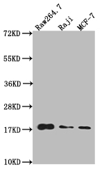
Western Blot
Positive WB detected in: Raw264.7 whole cell lysate, Raji whole cell lysate, MCF-7 whole cell lysate
All lanes: HistoneH3 antibody at 1:20000
Secondary
Goat polyclonal to mouse IgG at 1/50000 dilution
Predicted band size: 15-25 KDa
Observed band size: 15-25 KDa
Exposure time: 1min
-
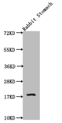
Western Blot
Positive WB detected in: Rabbit stomach tissue
All lanes: HistoneH3 antibody at 1:20000
Secondary
Goat polyclonal to mouse IgG at 1/50000 dilution
Predicted band size: 15-25 KDa
Observed band size: 15-25 KDa
Exposure time: 1min
-
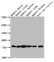
Western Blot
Positive WB detected in: Rabbit heart tissue, Rabbit liver tissue,Rabbit spleen tissue, Rabbit lung tissue, Rabbit kidney tissue, Rabbit small intestine tissue
All lanes: HistoneH3 antibody at 1:20000
Secondary
Goat polyclonal to mouse IgG at 1/50000 dilution
Predicted band size: 15-25 KDa
Observed band size: 15-25 KDa
Exposure time: 2min
-
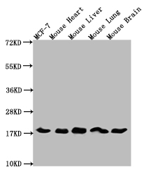
Western Blot
Positive WB detected in: MCF-7 whole cell lysate, Mouse heart tissue, Mouse liver tissue, Mouse lung tissue, Mouse brain tissue tissue
All lanes: HistoneH3 antibody at 1:5000
Secondary
Goat polyclonal to mouse IgG at 1/50000 dilution
Predicted band size: 15-25 KDa
Observed band size: 15-25 KDa
Exposure time: 5s
-
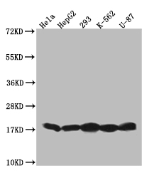
Western Blot
Positive WB detected in: Hela whole cell lysate, HepG2 whole cell lysate, 293 whole cell lysate, K562 whole cell lysate, U87 whole cell lysate
All lanes: HistoneH3 antibody at 1:20000
Secondary
Goat polyclonal to mouse IgG at 1/50000 dilution
Predicted band size: 15-25 KDa
Observed band size: 15-25 KDa
Exposure time: 2min
-
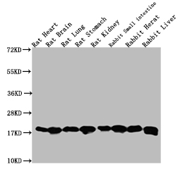
Western Blot
Positive WB detected in: Rat heart tissue, Rat brain tissue, Rat lung tissue, Rat stomach tissue, Rabbit small intestine tissue, Rabbit heart tissue, Rabbit liver tissue
All lanes: HistoneH3 antibody at 1:5000
Secondary
Goat polyclonal to mouse IgG at 1/50000 dilution
Predicted band size: 15-25 KDa
Observed band size: 15-25 KDa
Exposure time: 1min
-
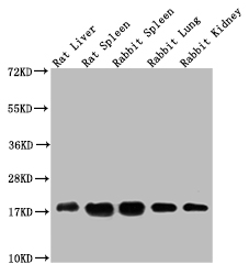
Western Blot
Positive WB detected in: Rat liver tissue, Rat spleen tissue, Rabbit spleen tissue, Rabbit lung tissue, Rabbit kidney tissue
All lanes: HistoneH3 antibody at 1:5000
Secondary
Goat polyclonal to mouse IgG at 1/50000 dilution
Predicted band size: 15-25 KDa
Observed band size: 15-25 KDa
Exposure time: 5s
-
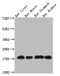
Western Blot
Positive WB detected in: Rat liver tissue, Rat brain tissue, Rat stomach tissue, Rat kidney tissue
All lanes: HistoneH3 antibody at 1:5000
Secondary
Goat polyclonal to mouse IgG at 1/50000 dilution
Predicted band size: 15-25 KDa
Observed band size: 15-25 KDa
-
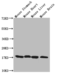
Western Blot
Positive WB detected in: Mouse stomach tissue, Mouse heart tissue, Mouse liver tissue, Mouse brain tissue
All lanes: HistoneH3 antibody at 1:20000
Secondary
Goat polyclonal to mouse IgG at 1/50000 dilution
Predicted band size: 15-25 KDa
Observed band size: 15-25 KDa
Exposure time: 2min
-
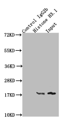
Immunoprecipitating HistoneH3 in K562 whole cell lysate
Lane 1: Mouse control IgG2b instead of CSB-MA010418A0m in Hela whole cell lysate.
Lane 2: CSB-MA010418A0m (2μg) + Hela whole cell lysate (500μg)
Lane 3: Hela whole cell lysate (20μg)
For western blotting, the blot was detected with CSB-MA010418A0m at 1:10000, and a HRP-conjugated Protein G antibody was used as the secondary antibody at 1:2000
-
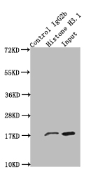
Immunoprecipitating HistoneH3 in Hela whole cell lysate
Lane 1: Mouse control IgG2b instead of CSB-MA010418A0m in Hela whole cell lysate.
Lane 2: CSB-MA010418A0m (2μg) + Hela whole cell lysate (500μg)
Lane 3: Hela whole cell lysate (20μg)
For western blotting, the blot was detected with CSB-MA010418A0m at 1:10000, and a HRP-conjugated Protein G antibody was used as the secondary antibody at 1:2000
-
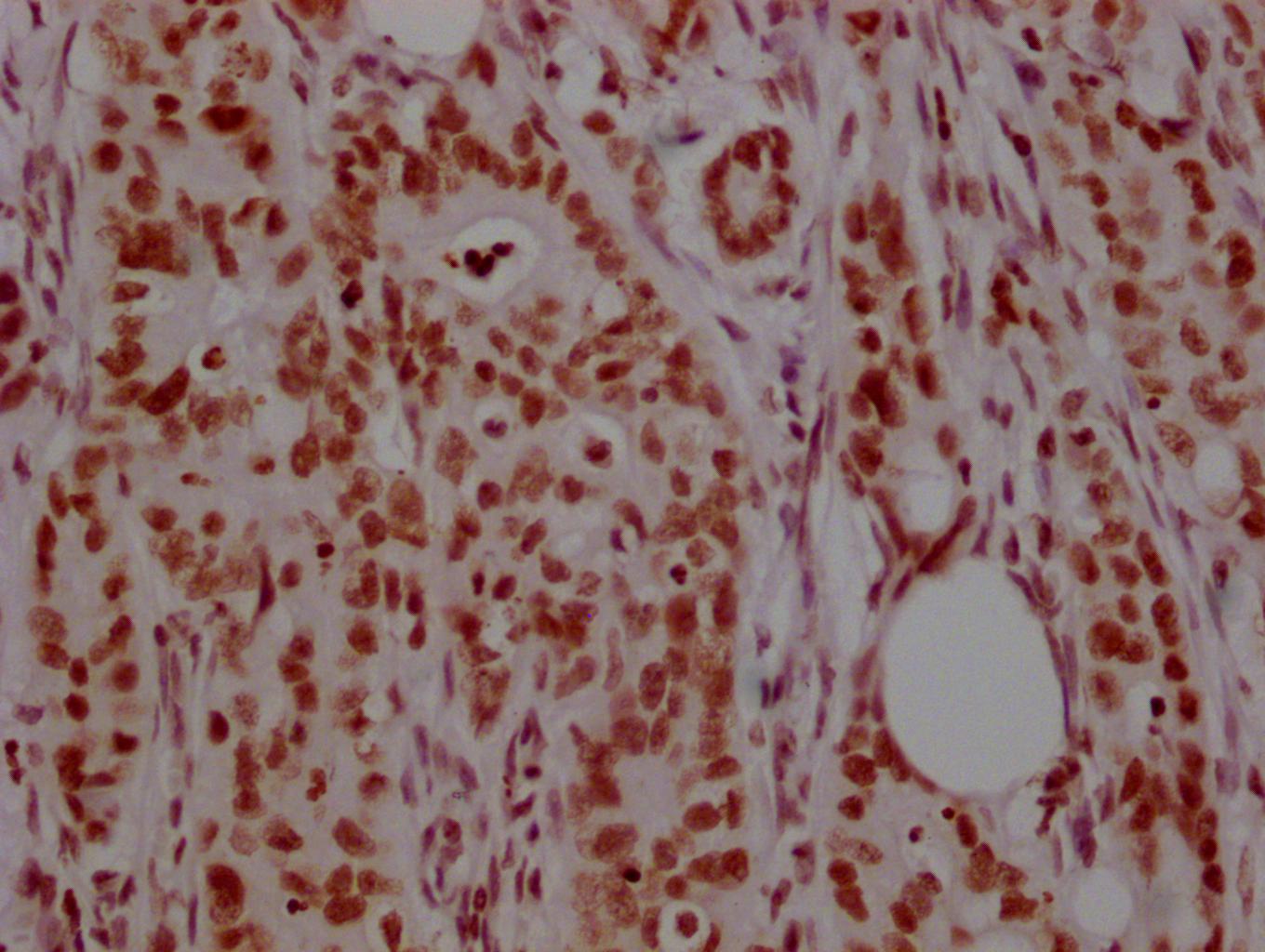
IHC image of CSB-MA010418A0m diluted at 1:200 and staining in paraffin-embedded human colon cancer performed on a Leica BondTM system. After dewaxing and hydration, antigen retrieval was mediated by high pressure in a citrate buffer (pH 6.0). Section was blocked with 10% normal goat serum 30min at RT. Then primary antibody (1% BSA) was incubated at 4°C overnight. The primary is detected by a biotinylated secondary antibody and visualized using an HRP conjugated SP system.
-
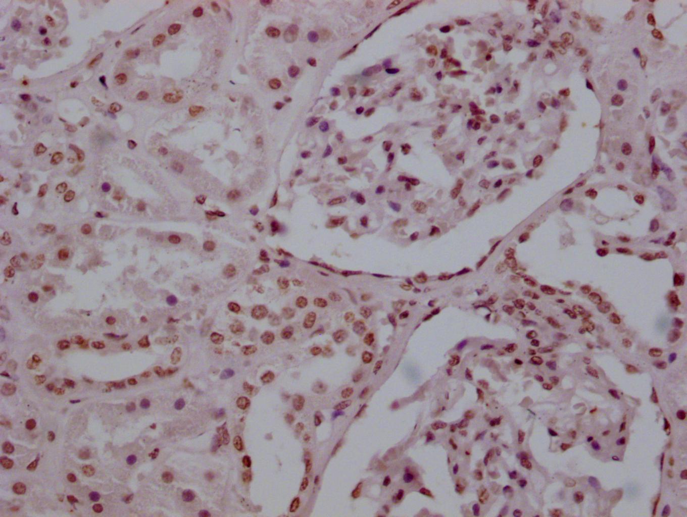
IHC image of CSB-MA010418A0m diluted at 1:200 and staining in paraffin-embedded human kidney tissue performed on a Leica BondTM system. After dewaxing and hydration, antigen retrieval was mediated by high pressure in a citrate buffer (pH 6.0). Section was blocked with 10% normal goat serum 30min at RT. Then primary antibody (1% BSA) was incubated at 4°C overnight. The primary is detected by a biotinylated secondary antibody and visualized using an HRP conjugated SP system.
-
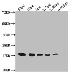
Western Blot
Positive WB detected in: Hela whole cell lysate at20μg, 10μg, 5μg, 2.5μg, 1.25μg, 0.625μg
All lanes: HistoneH3 antibody at 1:5000
Secondary
Goat polyclonal to mouse IgG at 1/50000 dilution
Predicted band size: 15-25 KDa
Observed band size: 15-25 KDa
Exposure time: 5s