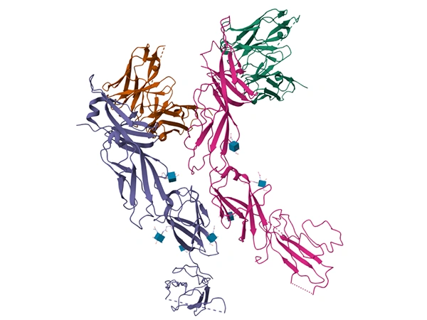
Lymphocyte-activation gene 3 (LAG3, CD223) is an important immune checkpoint molecule that was first cloned in 1990. Its gene shares approximately 20% homology with the CD4 gene, encoding a transmembrane protein containing 498 amino acids. This protein includes four immunoglobulin superfamily (IgSF) domains (D1-D4), and a unique cytoplasmic tail with the "KIEELE" motif and glutamate-proline (EP) repeat sequences, which are key to its immune regulatory functions [1].
Human LAG3 Structure (PDB 7TZG)
LAG3 is primarily expressed on the surface of immune cells such as activated T cells (CD4⁺, CD8⁺), regulatory T cells (Tregs), NK cells, B cells, and plasmacytoid dendritic cells (pDCs) [2,3]. In the tumor microenvironment, LAG3 is often co-expressed with other inhibitory receptors like PD1 on exhausted T cells. It inhibits T cell proliferation, cytokine secretion, and cytotoxic functions, thereby contributing to tumor immune evasion [1,3].
LAG3 can continuously associate with the T cell receptor (TCR)-CD3 complex and migrate to the immunological synapse (IS) after T cell activation. The EP repeat sequence in its cytoplasmic tail lowers the local pH at the immunological synapse, disrupting the zinc ion (Zn²⁺) binding between the CD4/CD8 co-receptors and the tyrosine kinase Lck. This leads to the dissociation of the co-receptor-Lck complex, ultimately inhibiting downstream TCR signaling (such as phosphorylation of ZAP70, ERK, and AKT), thereby restricting T cell activation. This process does not depend on its classical ligand, MHC class II molecules, and is the core mechanism by which LAG3 independently inhibits T cell function [3].
The main ligands of LAG3 include MHC class II molecules, fibrinogen-like protein 1 (FGL1), and liver sinusoidal endothelial cell lectin (LSECtin) [2]. The Loop 2 region (Gly¹⁰⁷-Pro¹¹⁵) in the D1 domain is the key interface for ligand binding, and binding to both MHC class II molecules and FGL1 depends on this region [4]. In addition, LAG3 forms homodimers through its D2 domain. The murine LAG3 dimer is mediated by interactions between Trp¹⁸⁰ and a hydrophobic pocket in the adjacent protomer, while the human LAG3 dimer relies on residues such as Phe²²⁷ and Trp¹⁸⁴. Dimerization enhances its binding capacity with ligands.
LAG3 and PD1 exhibit synergistic effects in T cell exhaustion. In chronic infection and tumor models, blocking either LAG3 or PD1 alone has limited effects on restoring T cell function, while combined blockade significantly reverses T cell exhaustion, enhancing cytokine secretion and proliferation. This synergy may be due to their distinct mechanisms of inhibiting TCR signaling: LAG3 primarily interferes with the co-receptor-Lck interaction, while PD1 inhibits downstream signaling through SHP-1/2 [1,3].
LAG3 mainly inhibits immune responses through the following pathways:
Abnormal regulation of IL2 can lead to immune imbalance and disordered signaling pathways, which are closely related to the occurrence and development of various diseases:
In 43 UM tissue samples, LAG3 expression was detected in 25 cases (58.1%), present in both tumor cells and tumor-infiltrating lymphocytes (TILs). In UM cases with high-risk histopathological factors (such as large cell type and necrosis), LAG3 gene expression was significantly upregulated. Patients with LAG3-positive TILs and high mRNA expression had significantly lower disease-free survival (DFS) (p<0.0001), suggesting that LAG3 can serve as a prognostic biomarker for high-risk UM patients. The mechanism may involve LAG3 inhibiting the antitumor activity of TILs, allowing tumor cells to evade immune surveillance [5].
Analysis of 61 GEP-NETs patients showed no LAG3-positive expression (0/61). However, other immune markers such as PD-L1 and MSI-H exhibited expression differences: PD-L1 had the highest positivity rate in G3 tumors (50%), and MSI-H was present in 80% of G1 tumors, with both more common in localized lesions. Although LAG3 was not positive in this tumor type, its co-absence with other immune checkpoints may contribute to immune evasion in GEP-NETs, with specific mechanisms requiring further research [6].
In breast cancer tissues, the presence of LAG3⁺ intraepithelial tumor-infiltrating lymphocytes (iTILs) is associated with improved breast cancer-specific survival (BCSS) (HR 0.71; 95% CI 0.56–0.90). Approximately 53% of PD-L1⁺ tumors and 61% of PD-1⁺ tumors had LAG3⁺ iTILs, and BCSS was better when LAG3⁺CD8⁺ iTILs co-infiltrated. This suggests that LAG3⁺ iTILs may reflect the body's antitumor immune activity, and their presence indicates a more effective immune response [7].
LAG3 is highly expressed on TILs in the NSCLC tumor microenvironment and is associated with tumor stage: patients with stage III had a higher proportion of LAG3⁺ cells in the tumor microcirculation than those with stage IV. LAG3 expression also correlated positively with PD-L1 expression. Patients with high LAG3 expression had lower response rates to PD-1 inhibitors and shorter progression-free survival (PFS), suggesting that LAG3 may be a marker of immune therapy resistance in NSCLC. The mechanism may involve LAG3 and PD1 synergistically inhibiting T cell function, weakening the effectiveness of immune therapy [8].
In patients with advanced gastric cancer treated with nivolumab monotherapy, the presence of LAG3⁺ TILs was associated with lower treatment response rates, and patients with high LAG3 mRNA levels had significantly shorter overall survival (OS). Additionally, LAG3 was negatively correlated with tumor mutation burden (TMB), suggesting that high LAG3 expression may inhibit T cell infiltration, reducing immune therapy sensitivity [9,10].
In locally advanced colorectal cancer with mismatch repair deficiency (MMR-deficient), neoadjuvant nivolumab combined with relatlimab (anti-LAG3) treatment induced significant pathological complete response (pCR), with increased CD8⁺ T cell infiltration and decreased LAG3⁺ cell proportion in the tumor microenvironment. This indicates that LAG3 in colorectal cancer promotes tumor progression by inhibiting cytotoxic T cell function, and blocking LAG3 can restore immune response [11].
Deficiency or dysfunction of LAG3 is associated with increased susceptibility to autoimmune diseases. In a type 1 diabetes model, Lag3⁻/⁻ mice had significantly increased numbers of CD4⁺ and CD8⁺ T cells infiltrating the islets, with accelerated disease progression. In patients with rheumatoid arthritis (RA), the proportion of LAG3⁺ Tregs was lower and negatively correlated with disease activity, suggesting that impaired immunosuppressive function of LAG3⁺ Tregs may exacerbate joint inflammation. The mechanisms mainly involve weakened inhibition of effector T cells by LAG3 and decreased Treg function, leading to excessive activation of autoreactive T cells [2].
In chronic hepatitis B virus (HBV) infection, LAG3⁺ erythroid progenitor cells inhibit HBsAg seroclearance by secreting TGF-β, and LAG3 expression levels correlate positively with HBsAg titers. The mechanism involves LAG3⁺ cells interacting with T cells through LAG3, while simultaneously releasing TGF-β to inhibit T cell clearance of HBV, delaying viral clearance [12].
The research progress on LAG3 (lymphocyte activation gene 3)-targeted drugs is rapid, with several drugs now in clinical trial stages. Among them, BMS-986016 (relatlimab), the first LAG3 blocker, has conducted multiple phase I to III clinical trials, showing good tolerability and antitumor activity. Other anti-LAG3 monoclonal antibodies such as LAG525, TSR-033, and INCAGN02385 are also in clinical trials, with preliminary data indicating that these drugs have good safety and potential antitumor effects. In addition, bispecific antibodies such as MGD013 (targeting both LAG3 and PD-1) have also shown good tolerability and antitumor activity. LAG3-targeted drugs have shown potential in both monotherapy and combination therapy, with some of the ongoing drug pipelines listed below:
Drug | Drug Type | Indications Under Investigation (Disease Names) | Research Institutions | Highest Development Stage |
---|---|---|---|---|
Nivolumab/Relatlimab | Monoclonal Antibody | Metastatic melanoma, unresectable melanoma, melanoma, tumor, metastatic colorectal cancer, metastatic microsatellite-stable colorectal cancer, etc. | Bristol Myers Squibb Co., Bristol-Myers Squibb Australia Pty Ltd., Bristol-Myers Squibb Pharma EEIG, Bristol-Myers Squibb (China) Investment Co., Ltd., Ono Pharmaceutical Co., Ltd., Shanghai Bristol-Myers Squibb Pharmaceuticals Co., Ltd. | Approved |
Fianlimab/Cemiplimab | Monoclonal Antibody | Unresectable melanoma, head and neck squamous cell carcinoma | Regeneron Pharmaceuticals, Inc. | Phase III |
Recombinant Human Lymphocyte Activation Gene-3 (hLAG-3) Fusion Protein (Immutep) | Fc Fusion Protein | Metastatic non-small cell lung cancer, metastatic HER2-negative breast cancer, HER2-positive metastatic breast cancer, soft tissue sarcoma, advanced head and neck squamous cell carcinoma, breast cancer, non-small cell lung cancer, head and neck squamous cell carcinoma, etc. | Immutep SAS, Taizhou Yibai Kang Pharmaceutical Technology Co., Ltd., Merck Sharp & Dohme LLC, Immutep Ltd., Taizhou Yiteng Jingang Pharmaceutical Co., Ltd. | Phase III |
Alcestobart | Monoclonal Antibody | Tumor, metastatic esophageal squamous cell carcinoma, esophageal squamous cell carcinoma, solid tumor, colorectal cancer, nasopharyngeal cancer, microsatellite-stable colorectal cancer, advanced cancer, advanced malignant solid tumor, etc. | Guangzhou BeiGene Biopharmaceutical Co., Ltd., Boqian Biotech Co., Ltd., BeiGene (Guangzhou) Pharmaceutical Co., Ltd., BeOne Medicines Ltd., Nanjing Weiling Bio-Technology Co., Ltd. | Phase III |
Relatlimab | Monoclonal Antibody | Recurrent non-squamous non-small cell lung cancer, unresectable melanoma, renal cell carcinoma, hepatocellular carcinoma, metastatic non-small cell lung cancer, etc. | Bristol Myers Squibb Co., Shanghai Bristol-Myers Squibb Pharmaceuticals Co., Ltd., Ono Pharmaceutical Co., Ltd. | Phase III |
Favezelimab/Pembrolizumab | Monoclonal Antibody | Colorectal adenocarcinoma, metastatic colorectal cancer, cutaneous squamous cell carcinoma, mismatch repair-deficient endometrial cancer, refractory classical Hodgkin lymphoma, etc. | Merck Sharp & Dohme Research (China) Co., Ltd., Merck Sharp & Dohme Corp., Merck Sharp & Dohme LLC, Merck & Co., Inc. | Phase III |
Fianlimab | Monoclonal Antibody | Melanoma, metastatic melanoma, advanced non-small cell lung cancer, locally advanced non-small cell lung cancer, muscle-invasive bladder cancer, etc. | Regeneron Pharmaceuticals, Inc., Memorial Sloan Kettering Cancer Center, Sanofi | Phase III |
Tepotinib | Bispecific Antibody | Gastroesophageal junction cancer, HER2-positive gastric cancer, metastatic HER2-positive gastroesophageal junction cancer | MacroGenics, Inc. | Phase II/III |
Nivolumab/Relatlimab/Hyaluronidase | Monoclonal Antibody | Metastatic melanoma | Bristol Myers Squibb Co. | Phase II |
FS-118 | Bispecific Antibody | Non-small cell lung cancer, diffuse large B cell lymphoma, head and neck tumor, hematologic tumor, colorectal cancer | Merck Serono SA, F-star Therapeutics, Inc., invoX Pharma Ltd., F-Star Delta Ltd., F Star Co., Ltd. | Phase II |
GSK-2831781 | Monoclonal Antibody | Ulcerative colitis, psoriasis | Immutep Ltd. | Phase II |
Tuparstobart | Monoclonal Antibody | Metastatic Merkel cell carcinoma, head and neck squamous cell carcinoma, metastatic melanoma, unresectable melanoma | Incyte Biosciences International SARL, Incyte Corp. | Phase II |
HLX-26 | Monoclonal Antibody | Advanced pulmonary non-squamous non-small cell lung cancer, advanced non-small cell lung cancer, metastatic non-small cell lung cancer, lymphoma, solid tumor, metastatic solid tumor, advanced malignant solid tumor, hepatocellular carcinoma | Shanghai Henlius Biotechnology Co., Ltd., Shanghai Fosun Pharmaceutical Industrial Development Co., Ltd., Shanghai Henlius Biopharmaceutical Co., Ltd. | Phase II |
DNV-3 | Monoclonal Antibody | Melanoma, esophageal squamous cell carcinoma, gastroesophageal junction cancer, hepatocellular carcinoma, lymphoma, small cell lung cancer, etc. | Zhejiang Timai Pharmaceutical Co., Ltd. | Phase II |
Tombestomig | Bispecific Antibody | PD-L1-positive triple-negative breast cancer, renal cell carcinoma, locally advanced renal cell carcinoma, locally advanced urothelial carcinoma, urothelial carcinoma, etc. | Hoffmann-La Roche, Inc., Roche Holding AG, F. Hoffmann-La Roche Ltd., Roche (China) Investment Co., Ltd. | Phase II |
Miptenalimab | Monoclonal Antibody | Esophageal adenocarcinoma, advanced cancer, advanced endometrial cancer, locally advanced unresectable gastric adenocarcinoma, melanoma, etc. | Boehringer Ingelheim GmbH | Phase II |
Cugrastomig | Bispecific Antibody | Colorectal adenocarcinoma, non-small cell lung cancer, non-small cell lung cancer stage IIIA, head and neck squamous cell carcinoma, etc., liver metastasis, melanoma, unresectable melanoma, refractory classical Hodgkin lymphoma | Zhongshan Kangfang Biomedical Co., Ltd., Kangfang Huike (Shanghai) Biotechnology Co., Ltd. | Phase I/II |
Fanastomig | Bispecific Antibody | Solid tumor | Anmab Biotechnology (Suzhou) Co., Ltd. | Phase I/II |
Recombinant Human Anti-LAG-3 Monoclonal Antibody (Guangzhou Yuheng Biotechnology/Wuxi AppTec) | Monoclonal Antibody | Advanced non-small cell lung cancer, advanced malignant solid tumor, melanoma | Guangzhou Yuheng Biotechnology Co., Ltd. | Phase I/II |
Favezelimab | Monoclonal Antibody | Hematologic tumor | Merck Sharp & Dohme LLC | Phase I/II |
TQB-2223/Penpulimab | Monoclonal Antibody | Advanced cancer | Zhengda Tianqing Pharmaceutical Group Co., Ltd. | Phase I |
AK137 | Bispecific Antibody | Advanced cancer | Zhongshan Kangfang Biomedical Co., Ltd. | Phase I |
INCA32459 | Bispecific Antibody | Metastatic head and neck squamous cell carcinoma, unresectable melanoma | Incyte Corp. | Phase I |
IBI-323 | Bispecific Antibody | Advanced cancer | Innovent Biologics (Suzhou) Co., Ltd. | Phase I |
IMP-761 | Monoclonal Antibody | Autoimmune diseases | Immutep Ltd. | Phase I |
ABL-501 | Bispecific Antibody | Advanced malignant solid tumor | ABL Bio, Inc. | Phase I |
Negalstobart | Monoclonal Antibody | Diffuse large B cell lymphoma, advanced cancer, gastroesophageal junction cancer, squamous non-small cell lung cancer, nasopharyngeal cancer | Innovent Biologics (Suzhou) Co., Ltd. | Phase I |
TQB-2223 | Monoclonal Antibody | Advanced hepatocellular carcinoma, advanced cancer, cholangiocarcinoma, ampullary cancer of Vater, gallbladder cancer, recurrent solid tumor, tumor metastasis | Symphogen A/S, Mingrui Pharmaceutical Co., Ltd., Zhengda Tianqing Pharmaceutical Group Co., Ltd., Clarusun Pharmaceutical Corp. | Phase I |
ZGGS15 | Bispecific Antibody | Advanced malignant solid tumor, solid tumor | Suzhou Zelgen Biopharmaceuticals Co., Ltd. | Phase I |
Recombinant Humanized Monoclonal Antibody MIL98 | Monoclonal Antibody | Advanced lymphoma, melanoma | Beijing Tiantan Bioscience Co., Ltd. | Phase I |
BJ-007 | Monoclonal Antibody | Infection | Shanghai BioJie Pharmaceutical Co., Ltd. | Clinical Application Approval |
(Data sourced from patsnap)
As a key immune checkpoint, LAG3 plays an important role in tumor immune evasion, autoimmune diseases, and chronic infections by interacting with the TCR-CD3 complex, interfering with co-receptor signaling, and binding to ligands. HUAMBI offers LAG3 recombinant proteins, antibodies, and ELISA kits to support your mechanism research or drug development.
● LAG3 Recombinant Protein
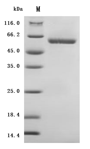
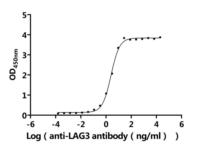
Recombinant Human Lymphocyte activation gene 3 protein (LAG3), partial (Active); CSB-MP012719HU3
References
[1] Nguyen L T, Ohashi P S. Clinical blockade of PD1 and LAG3--potential mechanisms of action[J]. Nat Rev Immunol, 2015, 15(1): 45-56.
[2] Hua S, et al. LAG3 (CD223) and autoimmunity: Emerging evidence[J]. J Autoimmun, 2020, 114: 102504.
[3] Guy C, et al. LAG3 associates with TCR-CD3 complexes and suppresses signaling by driving co-receptor-Lck dissociation[J]. Nat Immunol, 2022, 23(5): 757-767.
[4] Ming Q, et al. LAG3 ectodomain structure reveals functional interfaces for ligand and antibody recognition[J]. Nat Immunol, 2022, 23(7): 1031-1041.
[5] Singh L, et al. Prognostic significance of lymphocyte activation gene-3 (LAG3 gene) in uveal melanoma patients[J]. Annals of Oncology, 2021, 32(S7): S1463.
[6] Gurler F, et al. Retrospective analysis of PDL-1, LAG3, TIM3, OX40L and MSI status in gastroenteropancreatic neuroendocrine tumors (GEP-NETs)[J]. Annals of Oncology, 2021, 32(S5): S622.
[7] Burugu S, et al. LAG-3+ tumor infiltrating lymphocytes in breast cancer: clinical correlates and association with PD-1/PD-L1+ tumors[J]. Ann Oncol, 2017.
[8] LAG3 landscape in solid tumors and its association with immunotherapy outcomes in non-small cell lung cancer.
[9] LAG3-related factors to predict response to nivolumab monotherapy in advanced gastric cancer (WJOG10417GTR study).
[10] Phase 1 trial of the anti-LAG3 antibody favezelimab plus pembrolizumab in advanced gastric cancer.
[11] LBA31 Neoadjuvant nivolumab plus relatlimab (anti-LAG3) in locally advanced MMR-deficient colon cancers The NICHE-3 study.
[12] Xiu-Qing P, et al. LAG3+ erythroid progenitor cells inhibit HBsAg seroclearance during finite pegylated interferon treatment through LAG3 and TGF-β[J]. Antiviral Res, 2023.
Comments
Leave a Comment