Call us
301-363-4651 (Available 9 a.m. to 5 p.m. CST from Monday to Friday)
| Code | CSB-PA920084 |
| Size | US$166 |
| Order now | |
| Image |
|
| Have Questions? | Leave a Message or Start an on-line Chat |
| Application | Recommended Dilution |
|---|---|
| ELISA | 1:2000-1:5000 |
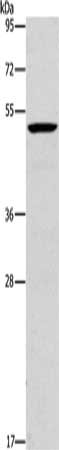
| WB | 1:500-1:2000 |
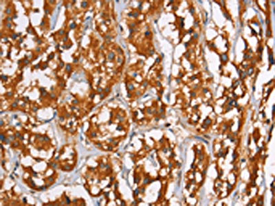
| IHC | 1:50-1:200 |
Applications : IF
Sample dilution: 1: 100
Review: the immunohistochemical staining for Plin2 protein 819 was more labelling in HF group, therefore the HF-EMPA presented less labelling, in second row.
By Anonymous
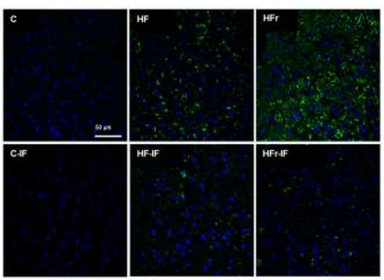
Applications : IF
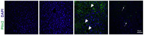
Review: Confocal laser scanning microscopy (blue: cell nuclei, DAPI; green: PLIN 2, Alexa 488). Abbreviations: C (control), IF (intermittent fasting), HF (high-fat), HFr (high-fructose).
By Anonymous
Applications : IF
Sample dilution: 1: 100
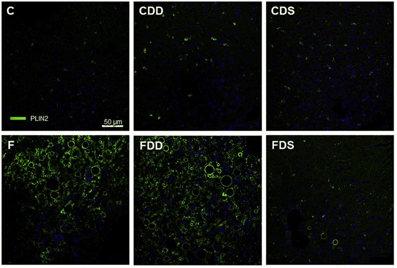
Review: Representative sections of the liver prepared for immunofluorescence with anti-Plin2 (green, confocal scanning laser microscopy to identify a constitutively associated cytoplasmic lipid droplet coat protein, same magnification in all photomicrographs). The identification of the groups is indicated in the left upper corner. There was increased Plin2 stain in CDD, and still more pronounced in F and FDD. In CDS and FDS the liver structure was restored.
By Anonymous
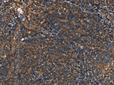
Applications : Immunofluorescence
Sample type: Mice
Sample dilution: 1:50
Review: The immunostaining for PLIN2 revealed the highest label was observed in the HF group (green label, white arrowheads), whereas the HF-L group presented with a significantly reduced label.
By Anonymous

