Full Product Name
Mouse anti-Homo sapiens (Human) CANX Monoclonal antibody
Alternative Names
Calnexin antibody; CALX_HUMAN antibody; CANX antibody; CNX antibody; FLJ26570 antibody; Histocompatibility complex class I antigen binding protein p88 antibody; IP90 antibody; Major histocompatibility complex class I antigen-binding protein p88 antibody; p90 antibody
Immunogen
Recombinant Human Calnexin protein (1-482AA)
Immunogen Species
Homo sapiens (Human)
Purification Method
>95%, Protein G purified
Concentration
It differs from different batches. Please contact us to confirm it.
Buffer
Preservative: 0.03% Proclin 300
Constituents: 50% Glycerol, 0.01M PBS, PH 7.4
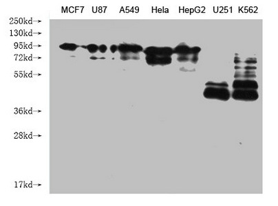
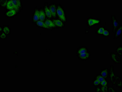
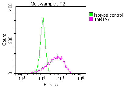
Tested Applications
ELISA, WB, IF, FC
Recommended Dilution
| Application |
Recommended Dilution |
| WB |
WB:1:1000-1:5000 |
| IF |
1:50-1:200 |
| FC |
1:50-1:200 |
Storage
Upon receipt, store at -20°C or -80°C. Avoid repeated freeze.
Lead Time
Basically, we can dispatch the products out in 1-3 working days after receiving your orders. Delivery time maybe differs from different purchasing way or location, please kindly consult your local distributors for specific delivery time.
Usage
For Research Use Only. Not for use in diagnostic or therapeutic procedures.