Call us
301-363-4651 (Available 9 a.m. to 5 p.m. CST from Monday to Friday)
| Code | CSB-PA000553 |
| Size | US$119 |
| Order now | |
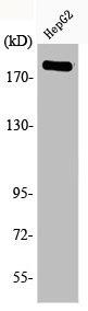
| Image |
|
| Have Questions? | Leave a Message or Start an on-line Chat |
| Application | Recommended Dilution |
|---|---|
| WB | 1:500-1:2000 |
| IHC | 1:100-1:300 |
| ELISA | 1:20000 |
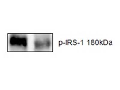
Applications : Western Blot (WB)
Sample type: mouse hippocampus
Sample dilution: 1:1000
Review: The samples of hippocampus were manually homogenized in a glass–glass potter containing ice-cold RIPA buffer in the presence of proteases and phosphatases inhibitor cocktail . The samples of hippocampus were diluted to a final protein concentration of 2 μg/μl in sample buffer. The samples (20 μg of protein) and prestained molecular weight standards were separated by 10% SDS-PAGE electrophoresis and transferred to nitrocellulose membrane (0.45 μm, Bio-Rad) using Transfer-Blot® Turbo™ Transfer System (1.0 A; 35 min) and equal protein loading was confirmed by Ponceau S staining. After blocking with 3% bovine serum albumin solution, the blots were incubated overnight at 4 °C with primary antibody. After the primary antibody incubations, membranes were washed and incubated for 1 h at room temperature with anti-rabbit secondary antibodies conjugated with horseradish peroxidase (1:5000, Bio-Rad Laboratories, Hercules, CA, USA). For protein detection, we used chemiluminescence kit (Amersham, SP, Brazil) and the signals were captured with Amersham Imager 600 (GE health care life sciences). The optical density (O.D.) of western blot bands was quantified using Image J (NIH, Bethesda, MD, USA) software for Windows. Each value was derived from the ratio between arbitrary units obtained by the protein band and the β-actin band. The results were expressed as ratio/β-actin.
By Bruna Weber Fulco

