-SDS.jpg)
-1.jpg)
-2.jpg)
Call us
301-363-4651 (Available 9 a.m. to 5 p.m. CST from Monday to Friday)
| Code | CSB-EP007717HU |
| Abbreviation | Recombinant Human EPCAM protein, partial |
| MSDS | |
| Size | $224 |
| Order now | |
| Image |
|
| Have Questions? | Leave a Message or Start an on-line Chat |
The recombinant human EPCAM protein tagged with an N-terminal 6xHis-SUMO is produced by cloning the EPCAM gene fragment (24-265aa) into an expression vector designed for E. coli systems. The N-terminal 6xHis-SUMO tag gene is also inserted into the vector. This recombinant vector is then introduced into E. coli and protein expression is induced using IPTG. Once expressed, the cells are lysed, and the EPCAM protein is captured using affinity chromatography. The purity of the recombinant EPCAM protein is assessed using SDS-PAGE, which confirms a high degree of purity, greater than 90%, making the protein ideal for experimental use.
The Human EPCAM, also known as CD326, is a type I transmembrane glycoprotein that plays a crucial role in cell adhesion, signaling, and various cellular processes such as proliferation and differentiation. It is primarily expressed in epithelial tissues and is particularly noted for its overexpression in various carcinomas, making it a significant marker in cancer research and therapy [1][2].
EPCAM is characterized by a large extracellular domain, a single transmembrane domain, and a short cytoplasmic domain. Beyond mere adhesion, EPCAM is also involved in intracellular signaling pathways that can influence cell behavior, including migration and differentiation [2][3].
EPCAM is often overexpressed in tumor cells compared to normal epithelial cells. Therapies targeting EPCAM have been developed to enhance the immune response against tumors expressing this molecule [4][5]. Additionally, its expression is associated with poor prognosis in various cancers, including breast and ovarian cancers, highlighting its potential as a biomarker for disease progression [1][3][6].
References:
[1] R. Ohashi, K. Kawahara, T. Fujii, H. Takei, & Z. Naito, Higher expression of epcam is associated with poor clinical and pathological responses in breast cancer patients undergoing neoadjuvant chemotherapy, Pathology International, vol. 66, no. 4, p. 210-217, 2016. https://doi.org/10.1111/pin.12404
[2] G. Carpenter and M. Brewer, Epcam: another surface-to-nucleus missile, Cancer Cell, vol. 15, no. 3, p. 165-166, 2009. https://doi.org/10.1016/j.ccr.2009.02.005
[3] M. Lee, Prognostic impact of epithelial cell adhesion molecule in ovarian cancer patients, Journal of Gynecologic Oncology, vol. 25, no. 4, p. 352, 2014. https://doi.org/10.3802/jgo.2014.25.4.352
[4] F. Suurs, G. Lorenczewski, S. Stienen, M. Friedrich, E. Vries, D. Groot, et al. The biodistribution of a cd3 and epcam bispecific t-cell engager is driven by the cd3 arm, Journal of Nuclear Medicine, vol. 61, no. 11, p. 1594-1601, 2020. https://doi.org/10.2967/jnumed.120.241877
[5] X. Zheng, X. Fan, B. Fu, M. Zheng, A. Zhang, K. Zhong, et al. Epcam inhibition sensitizes chemoresistant leukemia to immune surveillance, Cancer Research, vol. 77, no. 2, p. 482-493, 2017. https://doi.org/10.1158/0008-5472.can-16-0842
[6] S. Bellone, E. Siegel, E. Cocco, M. Cargnelutti, D. Silasi, M. Azodi, et al. Overexpression of epithelial cell adhesion molecule in primary, metastatic, and recurrent/chemotherapy-resistant epithelial ovarian cancer, International Journal of Gynecological Cancer, vol. 19, no. 5, p. 860-866, 2009. https://doi.org/10.1111/igc.0b013e3181a8331f
Applications : Control proteins
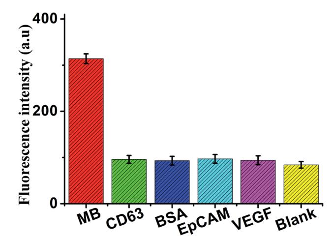
Review: Fluorescence intensity (at the emission wavelength of 517.6 nm) of the sensor in the presence of MB (5 ng mL -1 ), CD63 (50 ng mL -1 ), BSA (50 ng mL -1 ), EPCAM (50 ng mL -1 ), VEGF (50 ng mL -1 ) and black, respectively. Error bars: SD, n = 3.
By Anonymous
Applications : Drug related studies
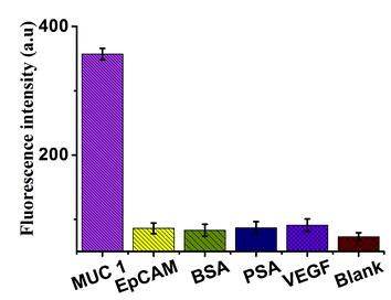
Review: Fluorescence intensity (at the emission wavelength of 519 nm) of the sensor in the presence of MUC1 (5 ng mL−1), EpCAM (50 ng mL−1), BSA (50 ng mL−1), PSA (50 ng mL−1), VEGF (50 ng mL−1) and black, respectively. In the presence of other control proteins (50 ng mL−1), the significant increase of fluorescence signal is observed in the presence of the MUC1 (5 ng mL−1), indicating that this proposed strategy exhibited good specificity for MUC1 detection.
By Anonymous
Applications : As control proteins
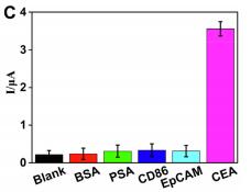
Review: To verify the specificity of the sensor for CEA detection, control experiments were carried out by using BSA, PSA, CD86 and EpCAM as control proteins. As shown in Fig. 4C, an obvious current was obtained in buffer containing CEA while only negligible currents were obtained in control groups containing BSA, PSA, CD86 and EpCAM even at a 10-fold concentration of CEA, indicating the specificity of the sensor.
By Anonymous
Applications : Protein-protein interaction
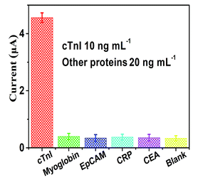
Review: there is no significant difference between the current response values of these four groups of proteins (myoglobin, EpCAM, CRP, and CEA) and the blank control group (the difference can be ignored). But he sensing platform has an excellent specificity towards the detection of cTnI.
By Anonymous

