Recombinant Mouse Platelet-derived growth factor D (Pdgfd)

In Stock
Code CSB-EP852889MO
Abbreviation Recombinant Mouse Pdgfd protein
MSDS
Size US$306
Order now
Image
  • (Tris-Glycine gel) Discontinuous SDS-PAGE (reduced) with 5% enrichment gel and 15% separation gel.

Have Questions? Leave a Message or Start an on-line Chat

Product Details

Purity
Greater than 90% as determined by SDS-PAGE.
Target Names
Uniprot No.
Research Area
Others
Alternative Names
Pdgfd; Scdgfb; Platelet-derived growth factor D; PDGF-D; Spinal cord-derived growth factor B; SCDGF-B) [Cleaved into: Platelet-derived growth factor D; latent form; PDGFD latent form); Platelet-derived growth factor D; receptor-binding form; PDGFD receptor-binding form)]
Species
Mus musculus (Mouse)
Source
E.coli
Expression Region
24-370aa
Target Protein Sequence
TPQRASIKALRNANLRRDESNHLTDLYQREENIQVTSNGHVQSPRFPNSYPRNLLLTWWLRSQEKTRIQLSFDHQFGLEEAENDICRYDFVEVEEVSESSTVVRGRWCGHKEIPPRITSRTNQIKITFKSDDYFVAKPGFKIYYSFVEDFQPEAASETNWESVTSSFSGVSYHSPSITDPTLTADALDKTVAEFDTVEDLLKHFNPVSWQDDLENLYLDTPHYRGRSYHDRKSKVDLDRLNDDVKRYSCTPRNHSVNLREELKLTNAVFFPRCLLVQRCGGNCGCGTVNWKSCTCSSGKTVKKYHEVLKFEPGHFKRRGKAKNMALVDIQLDHHERCDCICSSRPPR
Note: The complete sequence may include tag sequence, target protein sequence, linker sequence and extra sequence that is translated with the protein sequence for the purpose(s) of secretion, stability, solubility, etc.
If the exact amino acid sequence of this recombinant protein is critical to your application, please explicitly request the full and complete sequence of this protein before ordering.
Mol. Weight
56.2kDa
Protein Length
Full Length of Mature Protein
Tag Info
N-terminal 6xHis-SUMO-tagged
Form
Liquid or Lyophilized powder
Note: We will preferentially ship the format that we have in stock, however, if you have any special requirement for the format, please remark your requirement when placing the order, we will prepare according to your demand.
Buffer
If the delivery form is liquid, the default storage buffer is Tris/PBS-based buffer, 5%-50% glycerol.
Note: If you have any special requirement for the glycerol content, please remark when you place the order.
If the delivery form is lyophilized powder, the buffer before lyophilization is Tris/PBS-based buffer, 6% Trehalose.
Reconstitution
We recommend that this vial be briefly centrifuged prior to opening to bring the contents to the bottom. Please reconstitute protein in deionized sterile water to a concentration of 0.1-1.0 mg/mL.We recommend to add 5-50% of glycerol (final concentration) and aliquot for long-term storage at -20°C/-80°C. Our default final concentration of glycerol is 50%. Customers could use it as reference.
Troubleshooting and FAQs
Storage Condition
Store at -20°C/-80°C upon receipt, aliquoting is necessary for mutiple use. Avoid repeated freeze-thaw cycles.
Shelf Life
The shelf life is related to many factors, storage state, buffer ingredients, storage temperature and the stability of the protein itself.
Generally, the shelf life of liquid form is 6 months at -20°C/-80°C. The shelf life of lyophilized form is 12 months at -20°C/-80°C.
Lead Time
3-7 business days
Notes
Repeated freezing and thawing is not recommended. Store working aliquots at 4°C for up to one week.
Datasheet & COA
Please contact us to get it.
Description

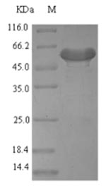
The recombinant mouse Pdgfd protein is co-expressed with an N-terminal 6xHis-SUMO tag in E. coli. The Pdgfd gene fragment (24-370aa) with the N-terminal 6xHis-SUMO tag gene is cloned into a suitable vector, which is transfected into E.coli cells. E.coli cells are induced to express the Pdgfd protein following the addition of IPTG. The Pdgfd protein is purified using Ni-NTA affinity chromatography. After elution, the purified recombinant Pdgfd protein is analyzed using SDS-PAGE to obtain a purity greater than 90%.

Mouse Pdgfd is secreted as a disulfide-bonded dimer and primarily signals through the PDGFR-β [1][2]. It has been implicated in several physiological and pathological conditions, including vascular remodeling, fibrosis, and tumor progression.

In the context of vascular biology, Pdgfd has been shown to induce significant changes in vascular smooth muscle cells (vSMCs), leading to conditions such as cardiac fibrosis and atherosclerosis [3][4]. Specifically, studies have demonstrated that overexpression of Pdgfd in transgenic mouse models results in enhanced proliferation of vSMCs, which contributes to the thickening of the vessel wall and subsequent cardiovascular complications [3][5]. Furthermore, Pdgfd has been associated with mesangial cell proliferation in renal glomerulopathy, indicating its role in kidney disease [6][7].

Pdgfd has been recognized for its involvement in tumor biology. It has been shown to promote tumor growth and invasion in various cancer models, including prostate and endometrial cancers [8][9]. In prostate cancer, Pdgfd not only enhances tumor cell proliferation but also facilitates interactions with the surrounding stroma, which is critical for tumor progression [10]. Additionally, Pdgfd has been linked to glioma pathogenesis, where it contributes to tumor growth and angiogenesis [11][12]. The signaling pathways activated by Pdgfd are complex and involve various downstream effects, including the activation of matrix metalloproteinases, which are crucial for extracellular matrix remodeling and tumor metastasis [9][13].

References:
[1] H. Gladh, E. Folestad, L. Muhl, M. Ehnman, P. Tannenberg, A. Lawrence, et al. Mice lacking platelet-derived growth factor d display a mild vascular phenotype, Plos One, vol. 11, no. 3, p. e0152276, 2016. https://doi.org/10.1371/journal.pone.0152276
[2] W. Huang and H. Kim, Dynamic regulation of platelet-derived growth factor d (pdgf-d) activity and extracellular spatial distribution by matriptase-mediated proteolysis, Journal of Biological Chemistry, vol. 290, no. 14, p. 9162-9170, 2015. https://doi.org/10.1074/jbc.m114.610865
[3] A. Pontén, E. Folestad, K. Pietras, & U. Eriksson, Platelet-derived growth factor d induces cardiac fibrosis and proliferation of vascular smooth muscle cells in heart-specific transgenic mice, Circulation Research, vol. 97, no. 10, p. 1036-1045, 2005. https://doi.org/10.1161/01.res.0000190590.31545.d4
[4] Z. Zhang, C. Ruan, J. Lin, L. Xu, X. Chen, Y. Du, et al. Perivascular adipose tissue–derived pdgf-d contributes to aortic aneurysm formation during obesity, Diabetes, vol. 67, no. 8, p. 1549-1560, 2018. https://doi.org/10.2337/db18-0098
[5] Y. Cheng, Z. Zhang, B. Lan, J. Lin, X. Chen, L. Kong, et al. Pdgf-d activation by macrophage-derived upa promotes angii-induced cardiac remodeling in obese mice, The Journal of Experimental Medicine, vol. 218, no. 9, 2021. https://doi.org/10.1084/jem.20210252
[6] K. Hudkins, D. Gilbertson, M. Carling, S. Taneda, S. Hughes, M. Holdren, et al. Exogenous pdgf-d is a potent mesangial cell mitogen and causes a severe mesangial proliferative glomerulopathy, Journal of the American Society of Nephrology, vol. 15, no. 2, p. 286-298, 2004. https://doi.org/10.1097/01.asn.0000108522.79652.63
[7] C. Roeyen, F. Eitner, P. Boor, M. Moeller, U. Raffetseder, L. Hanssen, et al. Induction of progressive glomerulonephritis by podocyte-specific overexpression of platelet-derived growth factor-d, Kidney International, vol. 80, no. 12, p. 1292-1305, 2011. https://doi.org/10.1038/ki.2011.278
[8] C. Ustach, M. Taube, N. Hurst, S. Bhagat, R. Bonfil, M. Cher, et al. A potential oncogenic activity of platelet-derived growth factor d in prostate cancer progression, Cancer Research, vol. 64, no. 5, p. 1722-1729, 2004. https://doi.org/10.1158/0008-5472.can-03-3047
[9] Y. Wang, H. Qiu, H. Weixu, S. Li, & J. Yu, Over-expression of platelet-derived growth factor-d promotes tumor growth and invasion in endometrial cancer, International Journal of Molecular Sciences, vol. 15, no. 3, p. 4780-4794, 2014. https://doi.org/10.3390/ijms15034780
[10] A. Najy, Y. Jung, J. Won, M. Conley-LaComb, A. Saliganan, C. Kim, et al. Cediranib inhibits both the intraosseous growth of pdgf d‐positive prostate cancer cells and the associated bone reaction, The Prostate, vol. 72, no. 12, p. 1328-1338, 2011. https://doi.org/10.1002/pros.22481
[11] A. Arias, M. Lamé, L. Santarelli, H. R, L. Greene, & J. Angelastro, Regulated atf5 loss-of-function in adult mice blocks formation and causes regression/eradication of gliomas, Oncogene, vol. 31, no. 6, p. 739-751, 2011. https://doi.org/10.1038/onc.2011.276
[12] P. Gong, Y. Wang, P. Ge, C. Bailey, P. Zhang, D. Zhang, et al. The hif1α-pdgfd-pdgfrα axis controls glioblastoma growth at normoxia/mild-hypoxia and confers sensitivity to targeted therapy by echinomycin, Journal of Experimental & Clinical Cancer Research, vol. 40, no. 1, 2021. https://doi.org/10.1186/s13046-021-02082-7
[13] L. Xu, R. Tong, D. Cochran, & R. Jain, Blocking platelet-derived growth factor-d/platelet-derived growth factor receptor β signaling inhibits human renal cell carcinoma progression in an orthotopic mouse model, Cancer Research, vol. 65, no. 13, p. 5711-5719, 2005. https://doi.org/10.1158/0008-5472.can-04-4313

Customer Reviews and Q&A

 Customer Reviews

There are currently no reviews for this product.

Submit a Review here

Target Background

Function
Growth factor that plays an essential role in the regulation of embryonic development, cell proliferation, cell migration, survival and chemotaxis. Potent mitogen for cells of mesenchymal origin. Plays an important role in wound healing. Has oncogenic potential and can induce tumor formation. Induces macrophage recruitment, increased interstitial pressure, and blood vessel maturation during angiogenesis. Can initiate events that lead to a mesangial proliferative glomerulonephritis, including influx of monocytes and macrophages and production of extracellular matrix.
Gene References into Functions
  1. Data (including data from studies using transgenic/mutant mice and cells from such mice) suggest that Pdgfd promotes cell proliferation, cell migration, and expression of inflammatory factors in adventitial fibroblasts; in obese mice, inhibition of Pdgfd significantly reduces aortic aneurysm formation (induced by infusion of angiotensin II). PMID: 29794241
  2. upregulated in kidney fibrosis, may mediate renal scarring, and is dispensable for normal kidney development and physiological functions PMID: 26924050
  3. Inactivation of the Pdgfd gene resulted in a mild phenotype in C57BL/6 mice, and the offspring was viable, fertile and generally in good health. PMID: 27032083
  4. PDGF-D intensifies fibrogenesis by interfering with the fibrolytic activity of the TIMP-1/MMP-2/MMP-9 system, and PDGF-D signaling is mediated through both PDGF-alpha and -beta receptors. PMID: 25576870
  5. Radial glia require PDGFD-PDGFRbeta signalling in human but not mouse neocortex PMID: 25391964
  6. PDGF D represents a potentially promising target for prostate carcinoma treatment resistance in the absence of PTEN function, and warrants further laboratory evaluation and clinical study. PMID: 24331662
  7. Data indicate that the proteolytic processing of full-length PDGF-D is required for PDGF-D activation of preosteoclasts, and that beta-PDGFR is present in activated osteoclasts. PMID: 22158043
  8. identified PDGF-DD as an important target gene for antiangiogenic therapy due to its pleiotropic effects on vascular and non-vascular cells. PDGF-DD inhibition may offer new therapeutic options to treat neovascular diseases PMID: 20231273
  9. Failure of laminin alpha4-mediated down-regulation of PDGF activity contributes to the progressive renal lesions in this animal model. PMID: 20035058
  10. These results suggest that PDGF-D induce cellular transformation and promote tumour growth by accelerating the proliferation rate of the tumour cells, and by stimulation of tumour neovascularization. PMID: 12629513
  11. splicing variant results in significant differences in peptide expression and function PMID: 12890490
  12. PDGF-D plays an important role in the pathogenesis of tubulointerstitial injury through binding of PDGF-Rbeta in obstructive ureteral nephropathy PMID: 14514732
  13. PDGF-DD, a novel mediator of smooth muscle cell phenotypic modulation, is upregulated in endothelial cells exposed to atherosclerosis-prone flow patterns. PMID: 19028801

Show More

Hide All

Subcellular Location
Secreted.
Protein Families
PDGF/VEGF growth factor family
Tissue Specificity
Expressed at high levels in developing heart, lung, kidney and some muscle derivatives. Moderately expressed in liver, brain and testis. In the kidney, localized to glomerular mesangial cells and vascular smooth muscle cells. Up-regulated in areas of rena
Database Links
icon of phone
Call us
301-363-4651 (Available 9 a.m. to 5 p.m. CST from Monday to Friday)
icon of address
Address
7505 Fannin St., Ste 610, Room 7 (CUBIO Innovation Center), Houston, TX 77054, USA
icon of social media
Join us with

Subscribe newsletter

Leave a message

* To protect against spam, please pass the CAPTCHA test below.
CAPTCHA verification
© 2007-2025 CUSABIO TECHNOLOGY LLC All rights reserved. 鄂ICP备15011166号-1
Place an order now

I. Product details

*
*
*
*

II. Contact details

*
*

III. Ship To

*
*
*
*
*
*
*

IV. Bill To

*
*
*
*
*
*
*
*