PSMA1 Recombinant Monoclonal Antibody

Code CSB-RA276081A0HU
Size US$210
Order now
Image
  • IHC image of CSB-RA276081A0HU diluted at 1:100 and staining in paraffin-embedded human colorectal cancer performed on a Leica BondTM system. After dewaxing and hydration, antigen retrieval was mediated by high pressure in a citrate buffer (pH 6.0). Section was blocked with 10% normal goat serum 30min at RT. Then primary antibody (1% BSA) was incubated at 4°C overnight. The primary is detected by a Goat anti-rabbit polymer IgG labeled by HRP and visualized using 0.05% DAB.
  • IHC image of CSB-RA276081A0HU diluted at 1:100 and staining in paraffin-embedded human small intestine tissue performed on a Leica BondTM system. After dewaxing and hydration, antigen retrieval was mediated by high pressure in a citrate buffer (pH 6.0). Section was blocked with 10% normal goat serum 30min at RT. Then primary antibody (1% BSA) was incubated at 4°C overnight. The primary is detected by a Goat anti-rabbit polymer IgG labeled by HRP and visualized using 0.05% DAB.
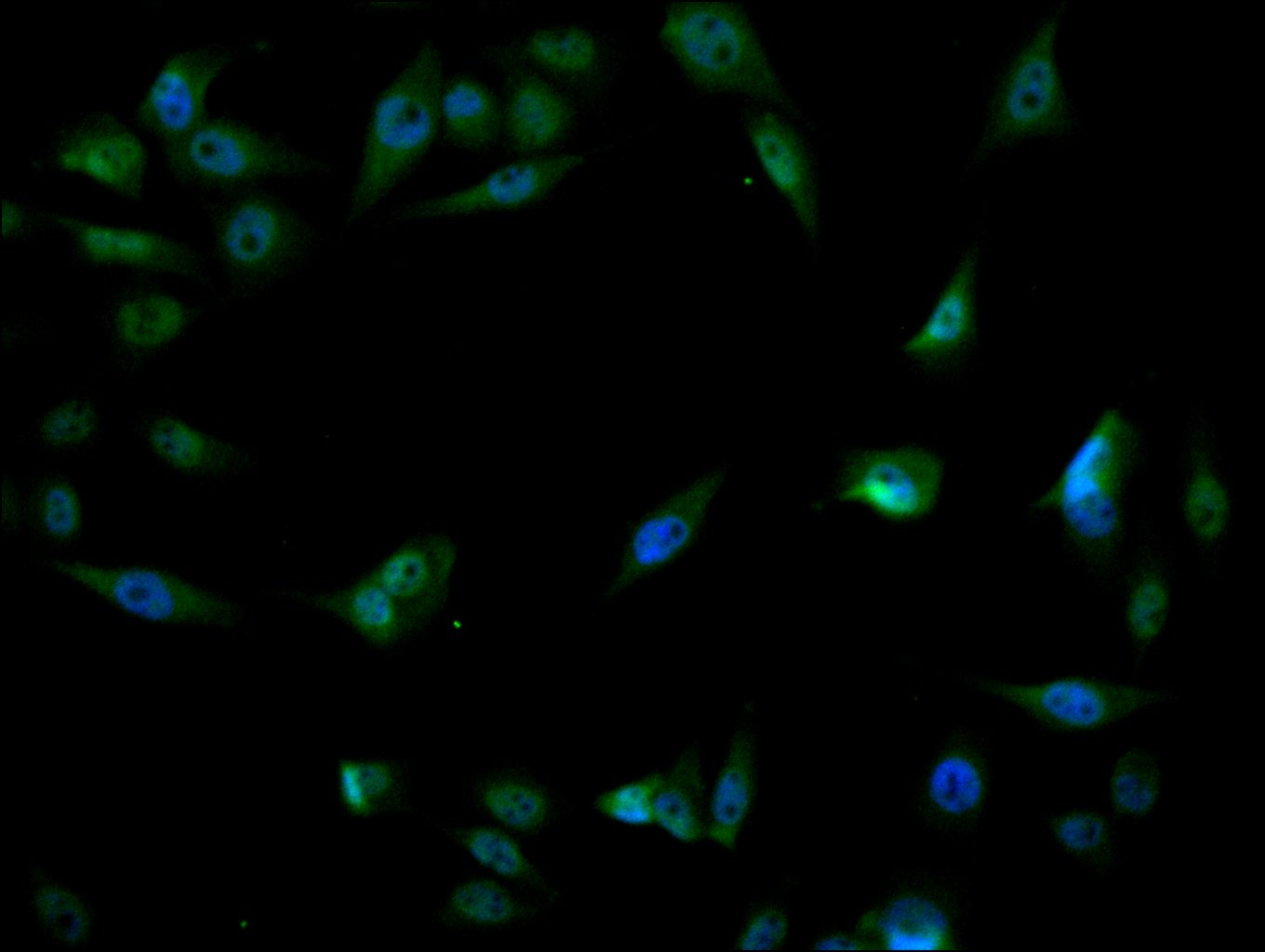
  • Immunofluorescence staining of PC-3 cell with CSB-RA276081A0HU at 1:50, counter-stained with DAPI. The cells were fixed in 4% formaldehyde, permeabilized using 0.2% Triton X-100 and blocked in 10% normal Goat Serum. The cells were then incubated with the antibody overnight at 4°C. The secondary antibody was Alexa Fluor 488-congugated AffiniPure Goat Anti-Rabbit IgG(H+L).
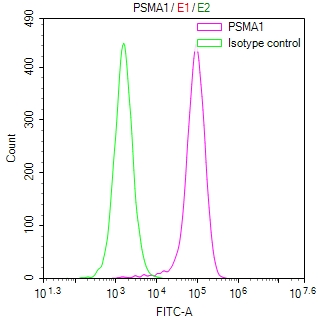
  • Overlay Peak curve showing HepG2 cells stained with CSB-RA276081A0HU (red line) at 1:100. The cells were fixed in 4% formaldehyde and permeated by 0.2% TritonX-100 for 10min. Then 10% normal goat serum to block non-specific protein-protein interactions followed by the antibody (1ug/1*106cells) for 45min at 4℃. The secondary antibody used was FITC-conjugated goat anti-rabbit IgG (H+L) at 1/200 dilution for 35min at 4℃.Control antibody (green line) was Rabbit IgG (1ug/1*106cells) used under the same conditions. Acquisition of >10,000 events was performed.
Have Questions? Leave a Message or Start an on-line Chat

Product Details

Uniprot No.
Target Names
Alternative Names
30 kDa prosomal protein antibody; HC 2 antibody; HC2 antibody; Macropain subunit C2 antibody; Macropain subunit nu antibody; MGC14542 antibody; MGC14575 antibody; MGC14751 antibody; MGC1667 antibody; MGC21459 antibody; MGC22853 antibody; MGC23915 antibody; Multicatalytic endopeptidase complex subunit C2 antibody; NU antibody; PROS 30 antibody; PROS-30 antibody; PROS30 antibody; Proteasome (prosome macropain) subunit alpha type 1 antibody; Proteasome alpha 1 subunit antibody; Proteasome component C2 antibody; Proteasome nu chain antibody; Proteasome subunit alpha type 1 antibody; Proteasome subunit alpha type I antibody; Proteasome subunit alpha type-1 antibody; Proteasome subunit nu antibody; Protein P30 33K antibody; PSA1_HUMAN antibody; PSC 2 antibody; PSC2 antibody; PSMA 1 antibody; psmA1 antibody
Species Reactivity
Human
Immunogen
A synthesized peptide derived from human PSMA1
Immunogen Species
Homo sapiens (Human)
Conjugate
Non-conjugated
Clonality
Monoclonal
Isotype
Rabbit IgG
Clone No.
16H4
Purification Method
Affinity-chromatography
Concentration
It differs from different batches. Please contact us to confirm it.
Buffer
Rabbit IgG in 10mM phosphate buffered saline , pH 7.4, 150mM sodium chloride, 0.05% BSA, 0.02% sodium azide and 50% glycerol.
Form
Liquid
Tested Applications
ELISA, IHC, IF, FC
Recommended Dilution
Application Recommended Dilution
IHC 1:50-1:200
IF 1:50-1:200
FC 1:50-1:200
Troubleshooting and FAQs
Storage
Upon receipt, store at -20°C or -80°C. Avoid repeated freeze.
Lead Time
Basically, we can dispatch the products out in 1-3 working days after receiving your orders. Delivery time maybe differs from different purchasing way or location, please kindly consult your local distributors for specific delivery time.
Description

PSMA1 (Proteasome subunit alpha type-1) serves as a core component of the 20S proteasome, the catalytic machinery responsible for regulated protein degradation in eukaryotic cells. This proteolytic complex plays essential roles in maintaining cellular homeostasis, regulating cell cycle progression, and modulating immune responses through antigen processing. Dysregulation of proteasome function has been implicated in numerous pathological conditions, including cancer and neurodegenerative diseases, making PSMA1 a valuable target for researchers investigating protein quality control mechanisms and therapeutic interventions.

This recombinant monoclonal antibody, clone 16H4, offers the reproducibility and consistency that demanding experimental workflows require. Generated against a synthetic peptide derived from human PSMA1 and produced using recombinant technology, this antibody provides sequence-defined specificity with minimal lot-to-lot variation, ensuring reliable results across extended studies and multi-site collaborations.

Validation data demonstrates robust performance across multiple detection platforms. Immunohistochemistry studies using human colorectal cancer and small intestine tissue sections show clear target detection at 1:100 dilution following citrate buffer antigen retrieval. Immunofluorescence analysis in PC-3 prostate cancer cells reveals distinct cytoplasmic staining patterns consistent with proteasome localization, while flow cytometry experiments using HepG2 hepatocellular carcinoma cells confirm specific signal separation from isotype controls. This versatility across tissue sections, adherent cell lines, and suspension-based assays provides researchers with flexibility in experimental design.

Whether investigating proteasome biology in cancer models, studying protein degradation pathways, or examining cellular stress responses, this antibody supports diverse research applications requiring reliable PSMA1 detection in human samples.

Usage
For Research Use Only. Not for use in diagnostic or therapeutic procedures.

Customer Reviews and Q&A

 Customer Reviews

There are currently no reviews for this product.

Submit a Review here

Target Background

Function
Component of the 20S core proteasome complex involved in the proteolytic degradation of most intracellular proteins. This complex plays numerous essential roles within the cell by associating with different regulatory particles. Associated with two 19S regulatory particles, forms the 26S proteasome and thus participates in the ATP-dependent degradation of ubiquitinated proteins. The 26S proteasome plays a key role in the maintenance of protein homeostasis by removing misfolded or damaged proteins that could impair cellular functions, and by removing proteins whose functions are no longer required. Associated with the PA200 or PA28, the 20S proteasome mediates ubiquitin-independent protein degradation. This type of proteolysis is required in several pathways including spermatogenesis (20S-PA200 complex) or generation of a subset of MHC class I-presented antigenic peptides (20S-PA28 complex).
Gene References into Functions
  1. In thyroid hemiagenesis, genomic examinations revealed the presence of four recurrent defects (three deletions and one duplication) affecting highly conservative proteasome genes PSMA1, PSMA3, and PSMD3. PMID: 28390009
  2. Data indicate that RNA interference screens identified PSMA1, a component of the 20S proteasome as target of non-small cell lung cancer (NSCLC) cytotoxicity. PMID: 24040035
  3. LC/MS/MS analysis revealed that the C-terminal 16 residues of sperm alpha6 subunit are processed. PMID: 21703233
Subcellular Location
Cytoplasm. Nucleus.
Protein Families
Peptidase T1A family
Database Links

HGNC: 9530

OMIM: 602854

KEGG: hsa:5682

STRING: 9606.ENSP00000414359

UniGene: Hs.102798

icon of phone
Call us
301-363-4651 (Available 9 a.m. to 5 p.m. CST from Monday to Friday)

icon of address
Address
No. 269, Shendun 5th Road, Donghu Hi-Tech Development Area, Hubei Province, 430206, P.R.China
icon of social media
Join us with

Subscribe newsletter

Leave a message

* To protect against spam, please pass the CAPTCHA test below.
CAPTCHA verification
© 2007-2026 CUSABIO TECHNOLOGY LLC All rights reserved. 鄂ICP备15011166号-1
Place an order now

I. Product details

*
*
*
*

II. Contact details

*
*

III. Ship To

*
*
*
*
*
*
*

IV. Bill To

*
*
*
*
*
*
*
*